
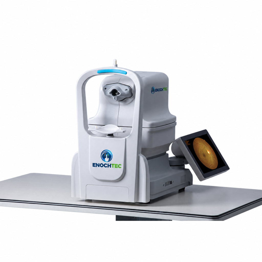
50° Fundus Imaging with Automatic and Manual Control
The YDC-3100 non-mydriatic fundus camera combines 5-megapixel photography with a 50° single-image field of view for professional retinal image documentation. Its acquisition modes include non-mydriatic and mydriatic photography, with automatic and manual focus, exposure and operation.
One-Key Workflow and Manual Adjustment
The automatic workflow supports chinrest adjustment, pupil searching, alignment, focusing and exposure, followed by sequential photography of both eyes through a one-key command. A dual-camera system assists autofocus and pupil location.
Manual operation is available when the automatic mode is not suitable or a particular area needs to be photographed. Touchscreen controls allow the operator to adjust alignment, focus, flash and diopter compensation.
Single Images and Nine-Position Mosaic
Each source image has a 50° field of view. The nine-position mosaic function combines nine 50° images into a composite view of up to 135°. This is a multi-image mosaic, not a 135° single exposure. Confirm mosaic software availability and the selected workflow in the quotation.
Pupil Size and Patient Preparation
The listed minimum pupil size is ≥2.8 mm, with a 13 mm working distance and ±25 D diopter compensation. Non-mydriatic capability does not guarantee that every patient can be imaged without dilation. Patient preparation and image assessment remain subject to the approved instructions for use and the responsible clinician’s judgement.
A soft-illumination workflow is also provided, intended to reduce illumination-related pupil constriction and permit two consecutive exposures. No quantitative illumination level, exposure interval or guaranteed patient response is specified; confirm the current operating instructions and selected configuration.
Image Processing and Data Integration
Image-processing controls include brightness, gamma, contrast and RGB adjustment. Lesion-area measurement and cup-to-disc-ratio tools are also provided; confirm their method, validation, diagnostic use and software version in the quotation. The system supports DICOM 3.0, PACS connection, real-time LAN sharing, USB storage expansion and FTP transfer. Confirm the required software, licences, network implementation, security and compatibility with the receiving system before ordering.
A customizable AI interface is listed. This is an integration option; it does not establish that an AI diagnostic system, algorithm, licence or regulatory clearance is included. Any AI-related scope requires a separate configuration review.
YDC-3100 Questions Before Ordering
Is the field of view 50° or 135°?
The single-image field of view is 50°. A mosaic of nine images can provide a composite view of up to 135°; it is not a single-shot 135° field.
Does the camera support manual operation?
Yes. Automatic and manual focus, exposure and operation are listed. Manual touchscreen adjustment covers alignment, focus, flash and diopter compensation.
Does every examination avoid dilation?
No. Both non-mydriatic and mydriatic acquisition modes are listed. The clinician determines the appropriate preparation and whether an image is adequate for the intended examination.
Are hospital integration and AI included?
DICOM 3.0 and a customizable AI interface are listed capabilities. The actual software, licences, receiving-system compatibility and implementation must be confirmed for the order; AI diagnosis is not assumed to be included.
Request a YDC-3100 Configuration
Contact ENOCHTEC sales with your country, quantity, intended examination setting and image-management requirements. Confirm the current model-specific specification, workstation, accessories, software, training, service and destination-market documentation in the quotation. Specifications and design are subject to change.


