
Solutions / Ophthalmology Clinic
Ophthalmology clinic planning
Ophthalmology Clinic Equipment Solution
Plan ophthalmology clinic equipment around the examinations your facility intends to provide. This guide connects examination, measurement, imaging and documentation modules with the decisions to resolve before model selection.
What equipment is used in an ophthalmology clinic?
An ophthalmology clinic equipment solution combines examination, measurement, imaging and documentation modules around the clinic’s intended services. It may include slit-lamp examination, tonometry, retinal imaging, visual-field testing, biometry or ultrasound, with refraction planned separately. The final configuration depends on clinical scope, room layout, patient flow, existing equipment and local requirements; it is not a fixed package.
Intended users
Start with rooms and examination scope.
This framework supports ophthalmology practices, outpatient departments, hospital project teams and distributors preparing a clinic shortlist. It can guide a new facility, an additional room or staged replacement without assuming that every clinic requires every category.
Recommended sequence
Plan around the clinic workflow.
Keep refraction, examination, imaging and specialist testing as defined modules. The exact procedure remains a matter for the qualified facility and applicable local requirements.
Services and rooms
List the examinations intended for each room and the users involved.
Anterior segment
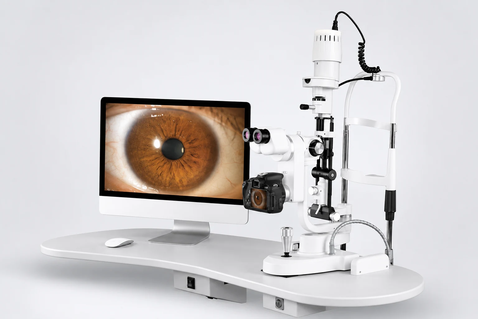
Choose the examination and documentation scope before selecting a slit-lamp configuration.
Pressure route
Compare tonometry approaches and verified operating requirements.
Retinal and functional modules
Add imaging or testing only where the intended service requires it.
Results and handoffs
Confirm room-to-room movement and any genuine device connections.
Typical equipment groups
Build the clinic in compatible modules.
The models below are examples to choose from, not a standard package. The product pages and the final quotation define the confirmed functions, specifications, included items and options.
Anterior-segment examination
Slit-lamp examination and documentation
Select a verified configuration that matches the examination and documentation scope. Confirm whether the selected item is a complete system, an imaging configuration or another device type.
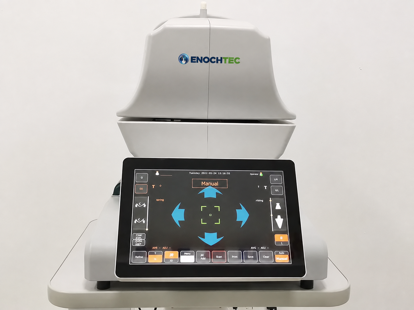
Intraocular-pressure measurement
Tonometry routes
Non-contact and rebound tonometers use different approaches. Compare positioning, room placement, probes or consumables and verified operating requirements.
Retinal imaging
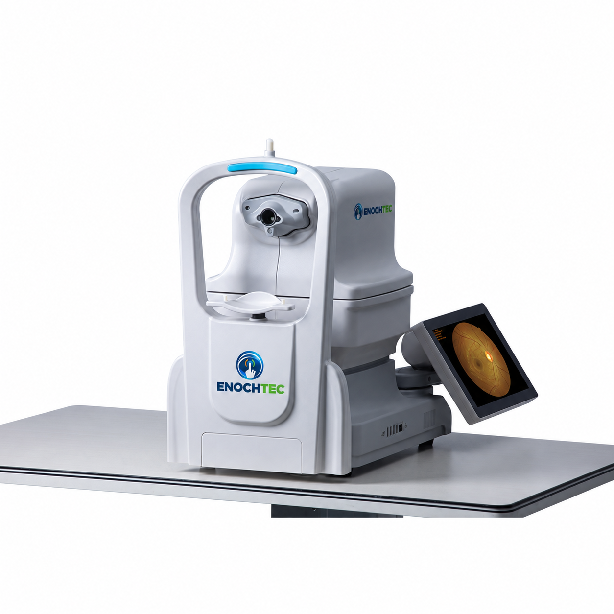
Fixed and handheld fundus photography
YDC-3100 is the current fixed-format non-mydriatic fundus-camera option. CFC-X is a handheld non-mydriatic fundus camera for portable retinal image capture. They serve different use formats and are not an interchangeable bundle. Detailed functions, accessories, software and any AI or DICOM capability are confirmed on the product page and in the quotation.
Functional testing
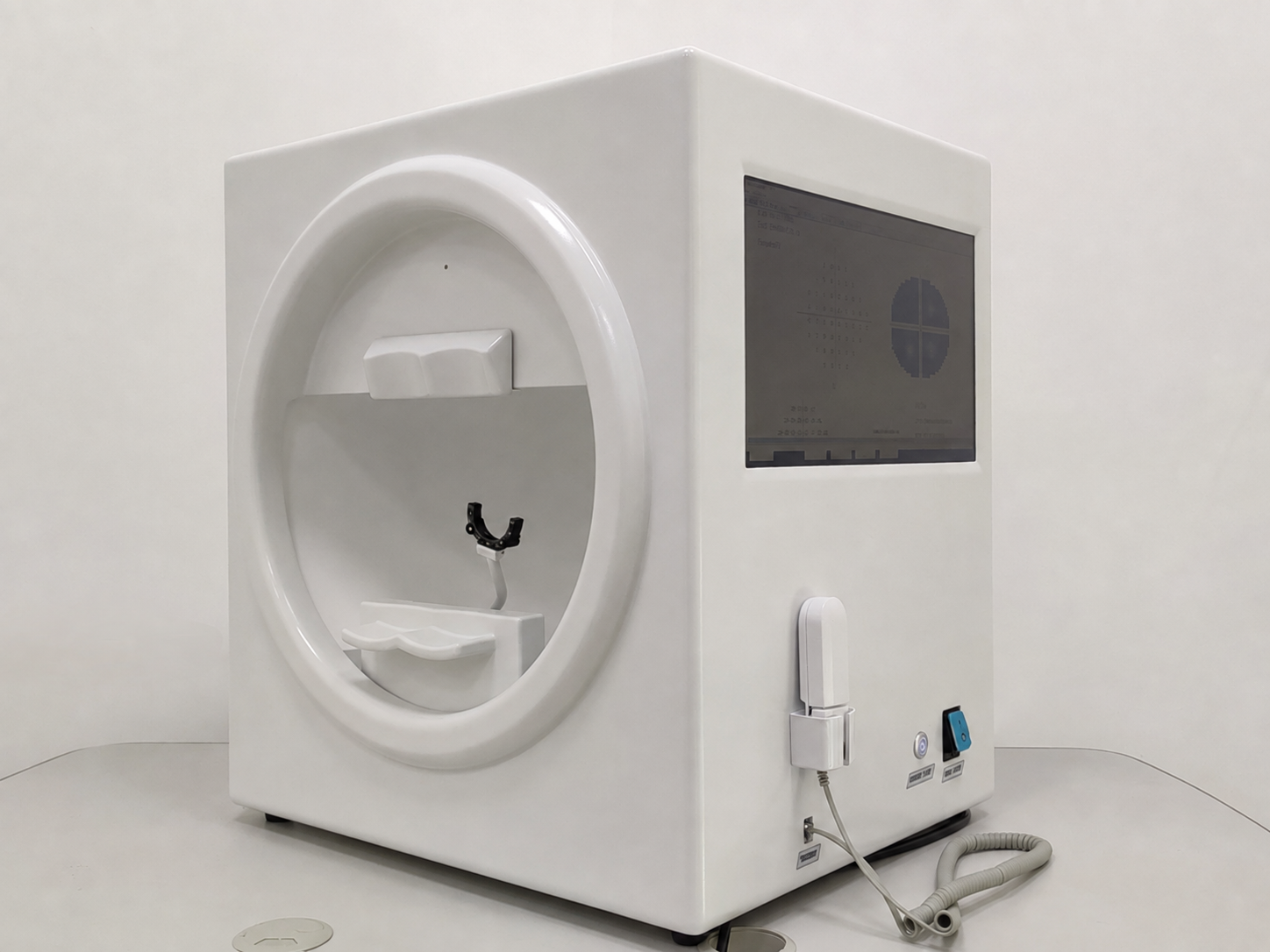
Visual-field assessment
BIO-1000 Plus is a computer perimeter. It should be considered only for the visual-field workflow and must not be confused with a biometer.
Biometry and ultrasound
Separate measurement groups
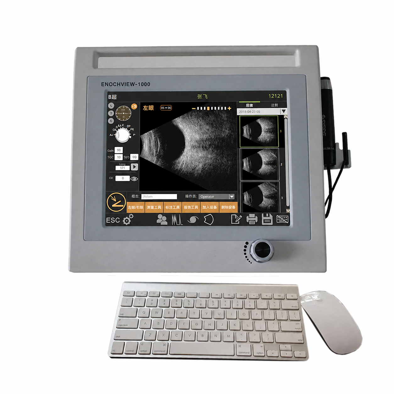
Optical biometry and ophthalmic A/B ultrasound serve different workflows. Include either module only where the intended pathway requires it.
Optional specialist modules
Screening and binocular vision
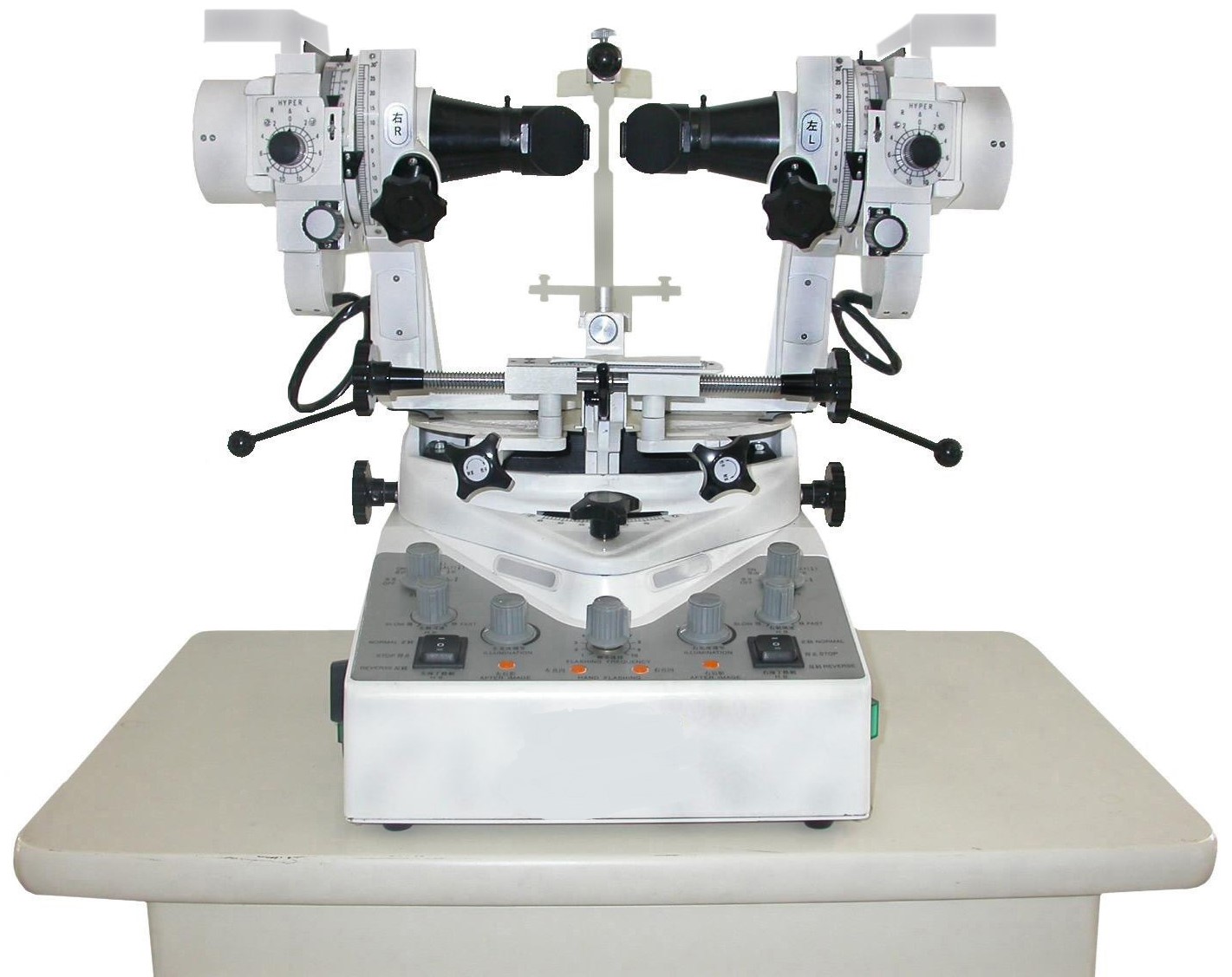
Screening and binocular-vision equipment are optional modules. Select them according to the intended workflow and the specifications on each model page.
Optional clinic air treatment
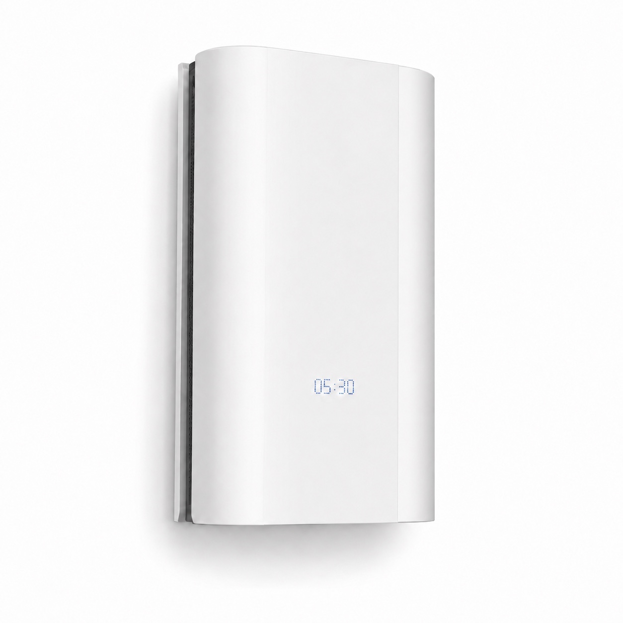
Review environmental air treatment separately
The BJ-1000 can be considered when a clinic brief includes a closed-loop UV air disinfector. It is optional environmental equipment, not part of the diagnostic workflow, and it does not replace ventilation, environmental cleaning, surface disinfection or instrument reprocessing.
Connected workflow
Refraction planning
Refraction may be part of the wider clinic, but a dedicated room should keep workstation, objective measurement, subjective refraction and visual-acuity decisions together.
Configuration choices
Resolve the scope before selecting models.
These decisions affect room allocation, placement, documentation and the final bill of materials.
Which examinations will each room provide?
Define current services and planned extensions before adding equipment groups.
Core or optional modules?
Separate essential workflow steps from later additions so a generic list does not become a mandatory package.
What equipment will remain?
Record existing devices and verify physical, electrical and data compatibility at model level.
What documentation workflow is required?
Confirm image, report and data-handling needs without assuming unverified integration.
Before quotation
Confirm every room and equipment group.
This page supports planning; the model pages and the final quotation define the actual configuration.
- Intended services and rooms
- Patient and operator flow
- Existing equipment
- Core and optional modules
- Dimensions, power and access
- Accessories and consumables
- Destination requirements
- Delivery and support scope
Buyer questions
Ophthalmology clinic FAQ.
Does every ophthalmology clinic need the same equipment?
No. The equipment set depends on the clinic’s services, number of rooms, users, patient flow, existing devices, budget and local requirements.
Is refraction equipment part of this solution?
Refraction may be part of the wider workflow, but a dedicated room should be planned through the Refraction Room Solution so its workstation and refraction decisions remain together.
Does a clinic need both a fundus camera and OCT?
Not automatically. They are different imaging categories and should be evaluated against the intended workflow, device functions, room plan and budget.
How should a buyer compare tonometer options?
Compare the measurement approach first, then verify positioning, probes or consumables, cleaning needs, dimensions, power and included items on each model page and in the quotation.
What information is needed for a configuration review?
Share intended services, room sizes, expected workflow, existing equipment, preferred equipment groups, destination market and any model preferences.
Build around the workflow


