
Examination and imaging
Ophthalmic Diagnostic & Imaging Equipment
Review equipment by examination need rather than treating every diagnostic device as interchangeable. Narrow the category first, then use each model page for available images and model-specific specifications.
What is ophthalmic diagnostic equipment?
Ophthalmic diagnostic equipment supports examination, measurement, screening or testing of the eye and visual system. Imaging is one part of that wider category. The exact function, output and included components must be confirmed on the relevant product page.
Organize by examination need
Keep each device type in its proper role.
The equipment types below help buyers distinguish imaging, measurement, screening and examination workflows before comparing individual products.
Retinal & Fundus Imaging
Equipment for fundus documentation and retinal imaging workflows.
Ultrasound & Biometry
Separate ophthalmic ultrasound from optical biometry and review the intended measurement workflow.
Slit Lamp Examination
Anterior-segment examination and imaging equipment, with included components confirmed by model.
Tonometry
Compare non-contact and rebound methods without treating them as technically identical.
Visual Field & Screening
Visual-field assessment and vision-screening equipment for their respective workflows.
Product models
Available product models
Use these model pages to review available images and configuration details. Each model page shows the specifications confirmed for that model.
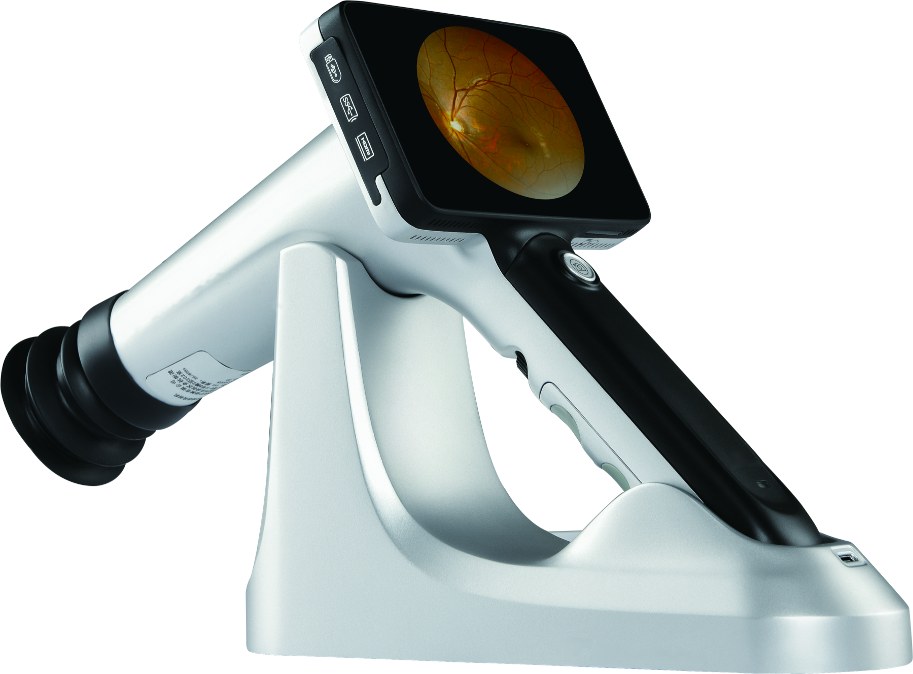 Portable retinal imaging
CFC-X Handheld Fundus Camera
Handheld non-mydriatic retinal camera for retinal image capture.
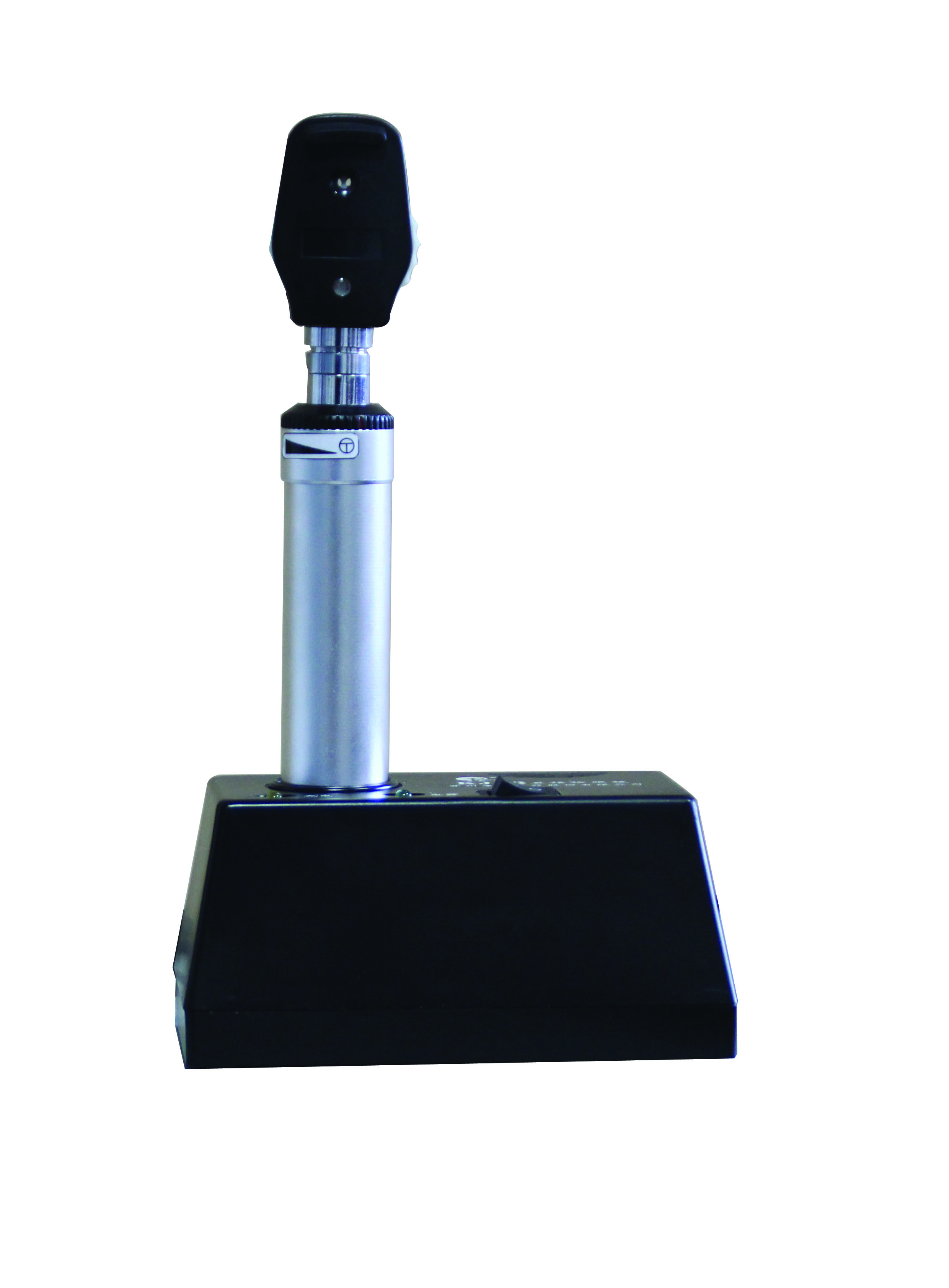 Fundus examination support
KJ8 Series Direct Ophthalmoscope
Direct ophthalmoscope for eye examination.
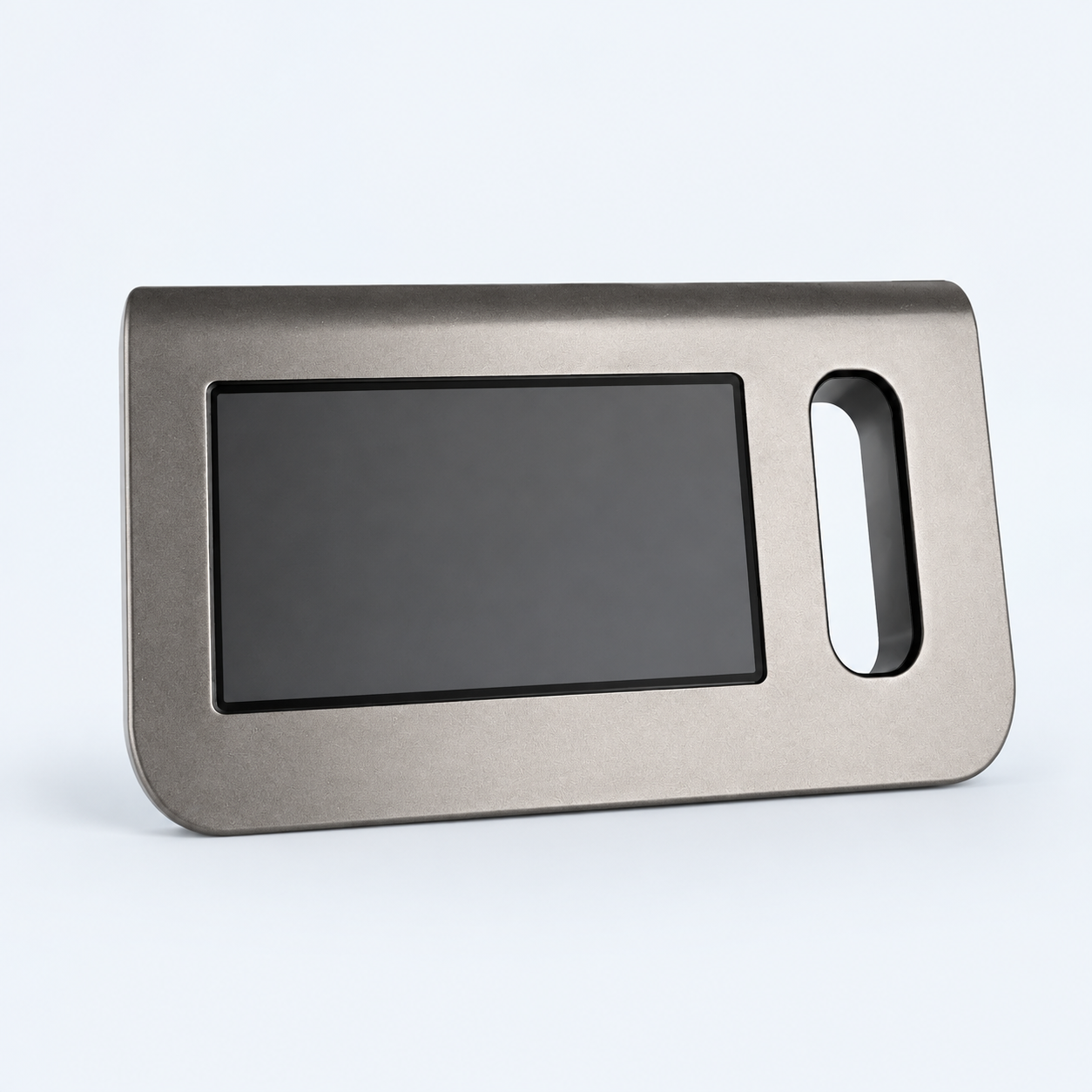 Product model
C10 Vision Screener
Binocular vision screener for preliminary refractive screening of both eyes.
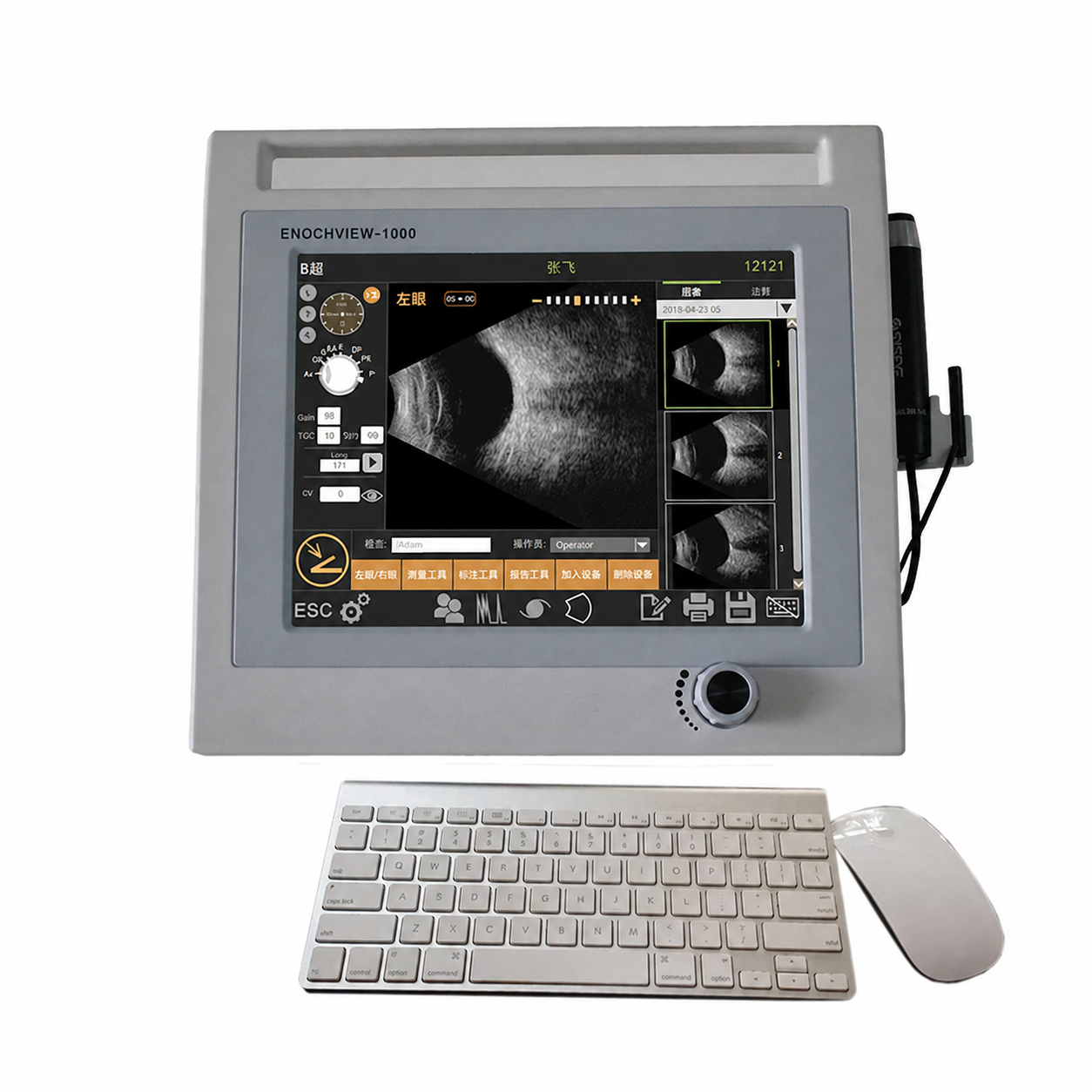 Product model
Enochview-1000 Ophthalmic A/B Ultrasound Scanner
Ophthalmic A/B ultrasound scanner for contact biometry and B-scan imaging.
 Product model
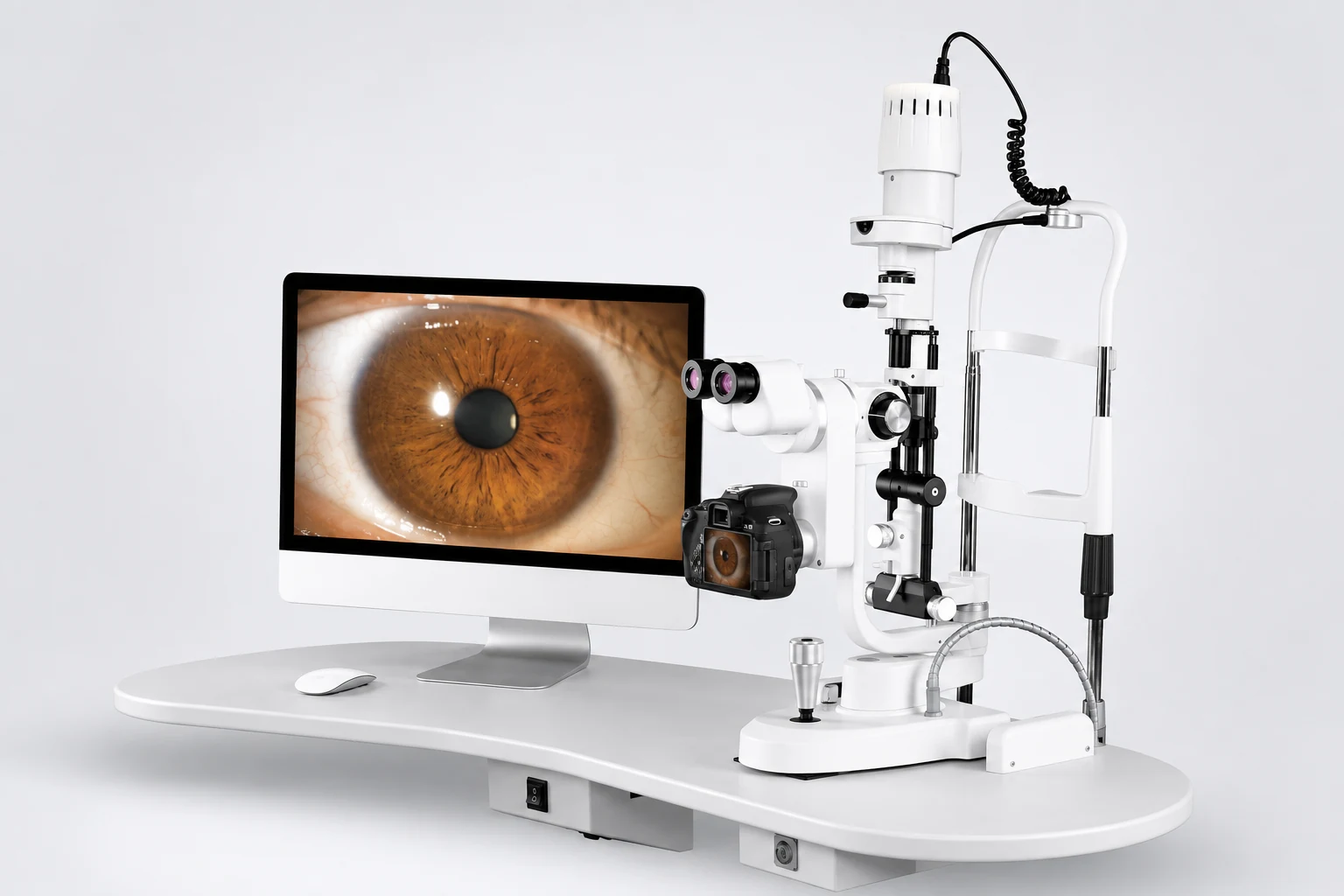
ML-350 / ML-350A Slit Lamp Microscope
Slit lamp for anterior-segment examination, with optional digital imaging.
 Product model
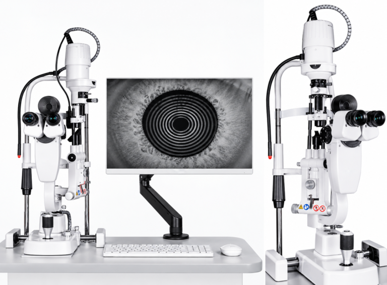
SLR-3000 Dry Eye Analyzer Slit Lamp
Slit-lamp system for tear-film assessment, meibography and blink analysis.
 Product model
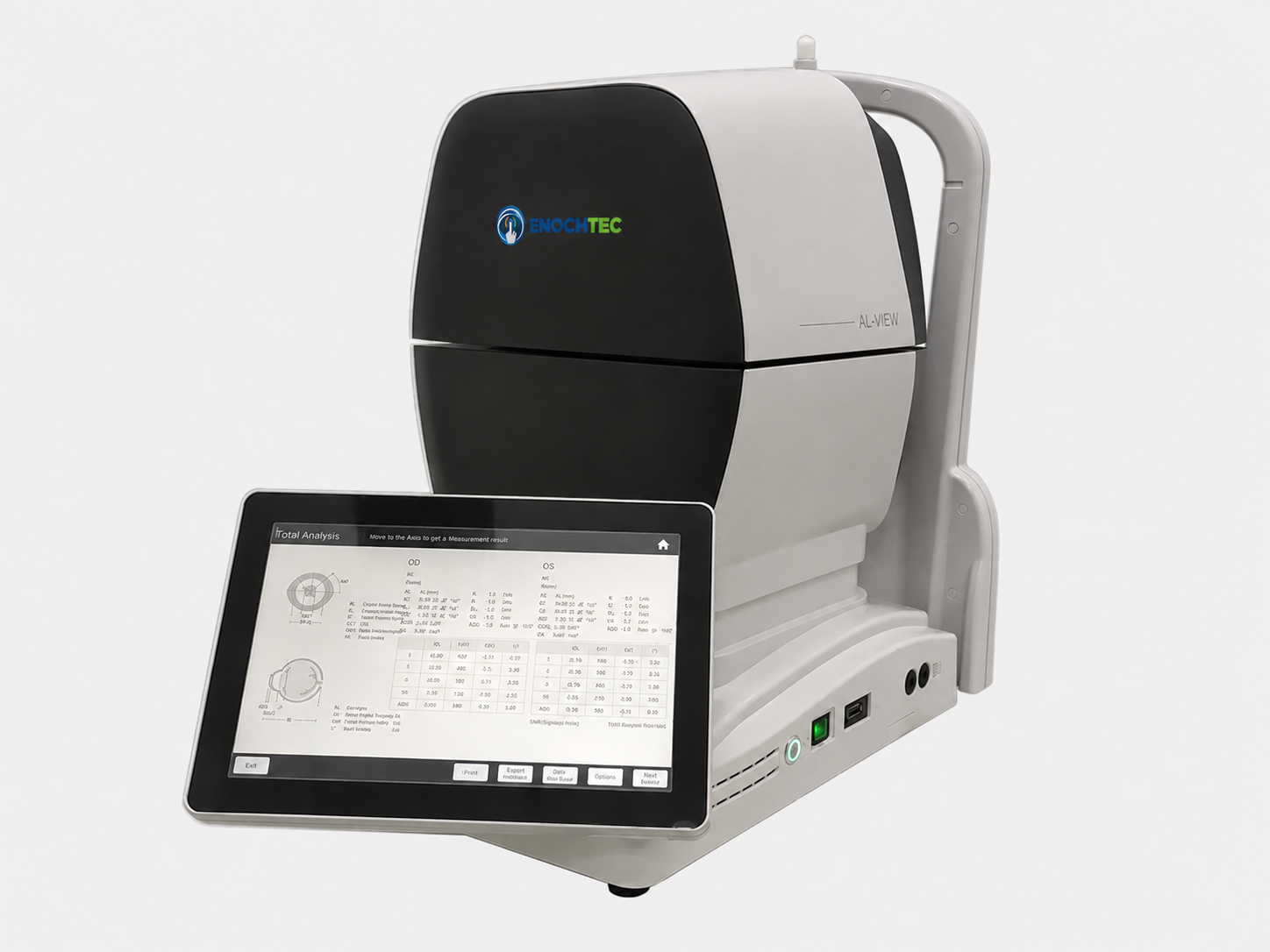
AL-VIEW Lite Optical Biometer
Optical biometer for measuring ocular parameters, including axial length.
 Product model
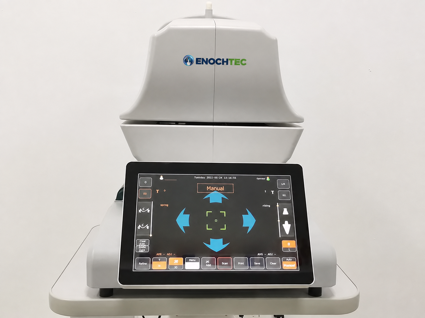
ST-1000P Non-contact Tonometer
Non-contact tonometer for professional measurement of intraocular pressure.
 Product model
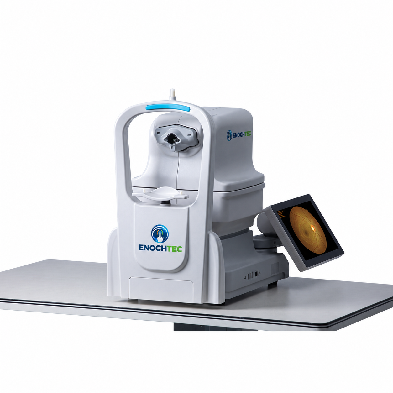
YDC-3100 Non-Mydriatic Fundus Camera
Non-mydriatic fundus camera for retinal image capture and documentation.
 Product model
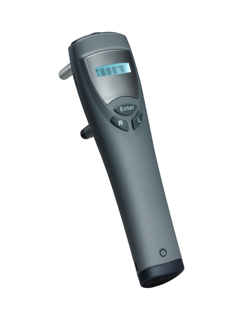
RT-500 Rebound Tonometer
Handheld rebound tonometer for professional measurement of intraocular pressure.
 Product model
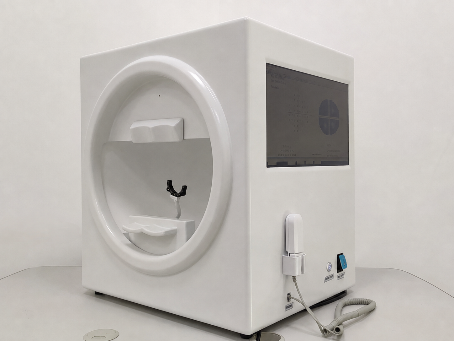
BIO-1000 Plus Computer Perimeter
Automatic static perimeter for professional visual-field assessment.
 Digital slit lamp
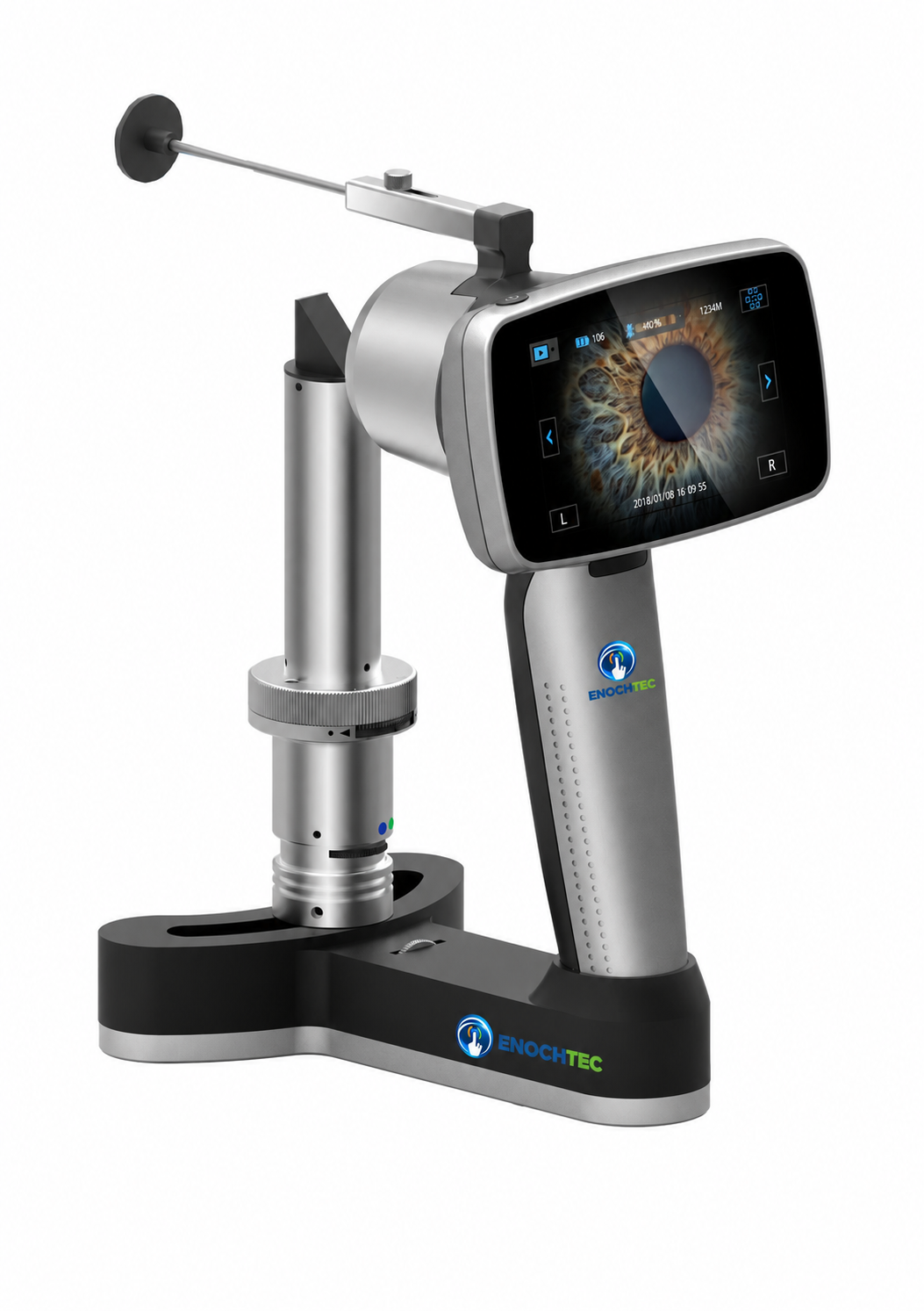
PSL-4961 Digital Hand-held Slit Lamp
Portable digital slit lamp for anterior-segment examination and image documentation.
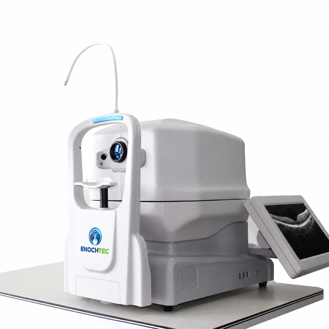 Optical coherence tomography
OCT-500 Spectral-Domain OCT System
Spectral-domain OCT for retinal and anterior-segment imaging.
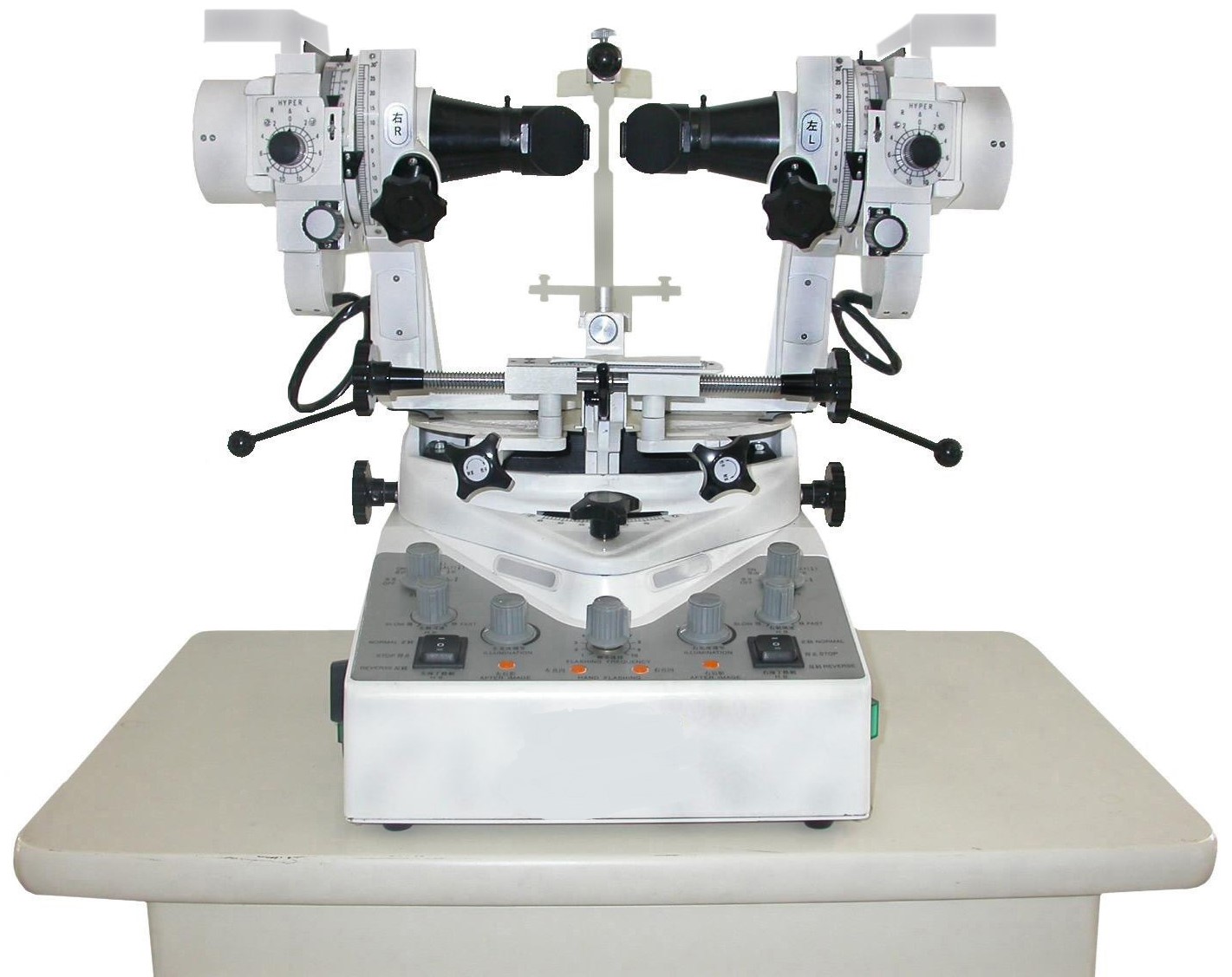 Binocular vision assessment
TS-23N Synoptophore Machine
Synoptophore for binocular-vision assessment and orthoptic training.
Selection guidance
What should be confirmed before a quotation?
Use the intended clinical workflow and required output to build the shortlist; do not assume the same bundle or software across models.
- Examination purpose — State the measurement, image, screening or test that the equipment must support.
- Required output — Clarify whether images, measurements, reports or other records are required.
- Included components — Confirm computers, cameras, software, accessories and optional modules in writing.
- Installation context — Provide room conditions, quantity, destination and existing equipment information.
From need to shortlist
From examination need to a confirmed configuration
A short workflow brief prevents a category label from being mistaken for a complete product specification.
Define the examination
Identify the clinical or screening task.
Choose the device type
Use the relevant category or equipment family.
Check the model page
Review specifications and configuration details.
Confirm the scope
Record included items, options and support at quotation.
Buyer questions
Answers before model selection.
What is the difference between diagnostic and imaging equipment?
Diagnostic equipment is the wider group and can include measurement, examination, screening and testing devices. Imaging equipment records or displays visual information.
Does every system include a computer, camera or software?
No common bundle should be assumed. Included components and optional modules vary by model and quotation.
How should I choose between tonometer types?
Start with the intended workflow and environment, then compare the method, accessories and supported use of the specific models.
Can these devices be planned as part of a clinic project?
Yes. Use the Ophthalmology Clinic or Hospital & Project Solution to organize rooms and workflows, then confirm every model separately.
Prepare the right shortlist


