
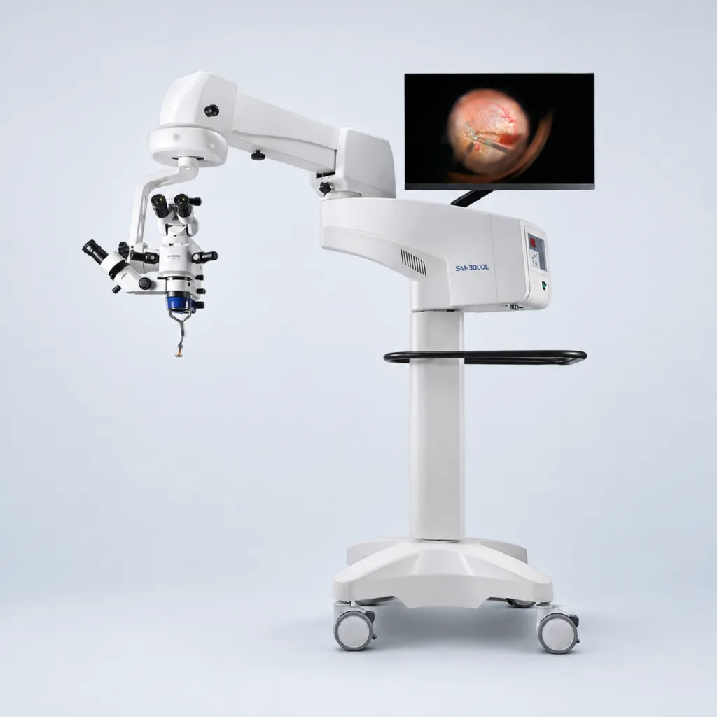
Key features
Configured for ophthalmic microsurgery.
Stereo viewing, foot-controlled adjustment and flexible positioning on one surgical microscope platform.
Anterior and posterior segment surgery
Configure the SM-3000L for anterior-segment procedures and extend the setup for posterior-segment surgery with the appropriate indirect viewing attachment and optical inverter.
Olympus achromatic optics
Olympus optics and an achromatic optical system provide a detailed stereoscopic working view, with an 8–50 mm field of view and a 200 mm working distance.
4–28× motorized zoom
Adjust continuous 4–28× magnification, X-Y positioning and motorized focus from the foot panel, keeping essential microscope movements within easy reach.
Coaxial stereo assistant viewing
The coaxial stereo assistant microscope offers 6×, 10× and 16× magnification. The assistant assembly can be placed on the left or right to suit the room layout.
Flexible binocular viewing
Folding 0–180° binoculars and a choice of 10× or 12× wide-field eyepieces help the surgeon set up the working position and view.
Optics & positioning
A working view, configured around the surgeon.
Olympus optics, motorized adjustment and stereo assistant viewing come together on an articulated floor stand.
A detailed view, from wide field to close focus.
The achromatic optical system combines 4–28× electric continuous zoom with an 8–50 mm field of view and a 200 mm working distance. Folding 0–180° binoculars, adjustable diopters and a 55–75 mm interpupillary range let the surgeon set up the viewing position.
Stereo viewing for the assistant.
The stereo coaxial assistant microscope offers 6×, 10× and 16× magnification, with zoom and focus functions. The assistant assembly can be positioned on the left or right to suit the operating-room layout.
LED illumination, adjusted to the view.
The aspheric LED system provides at least 80,000 lux, with eight brightness levels available through the foot control and control panel. An adjustable spot diameter, adjustable red reflex and macular-protection, green and cobalt-blue filters support the selected viewing setup.
Reach and positioning for the operating room.
A 1360 mm arm extension and ±250 mm vertical adjustment provide positioning flexibility around the operating table. Electromagnetic joint locks and dual-handle operation control the stand, supported by a 720 × 720 mm base.
A closer look
Foot control and posterior-segment accessories.
Explore the control layout and the optional viewing components for the SM-3000L.
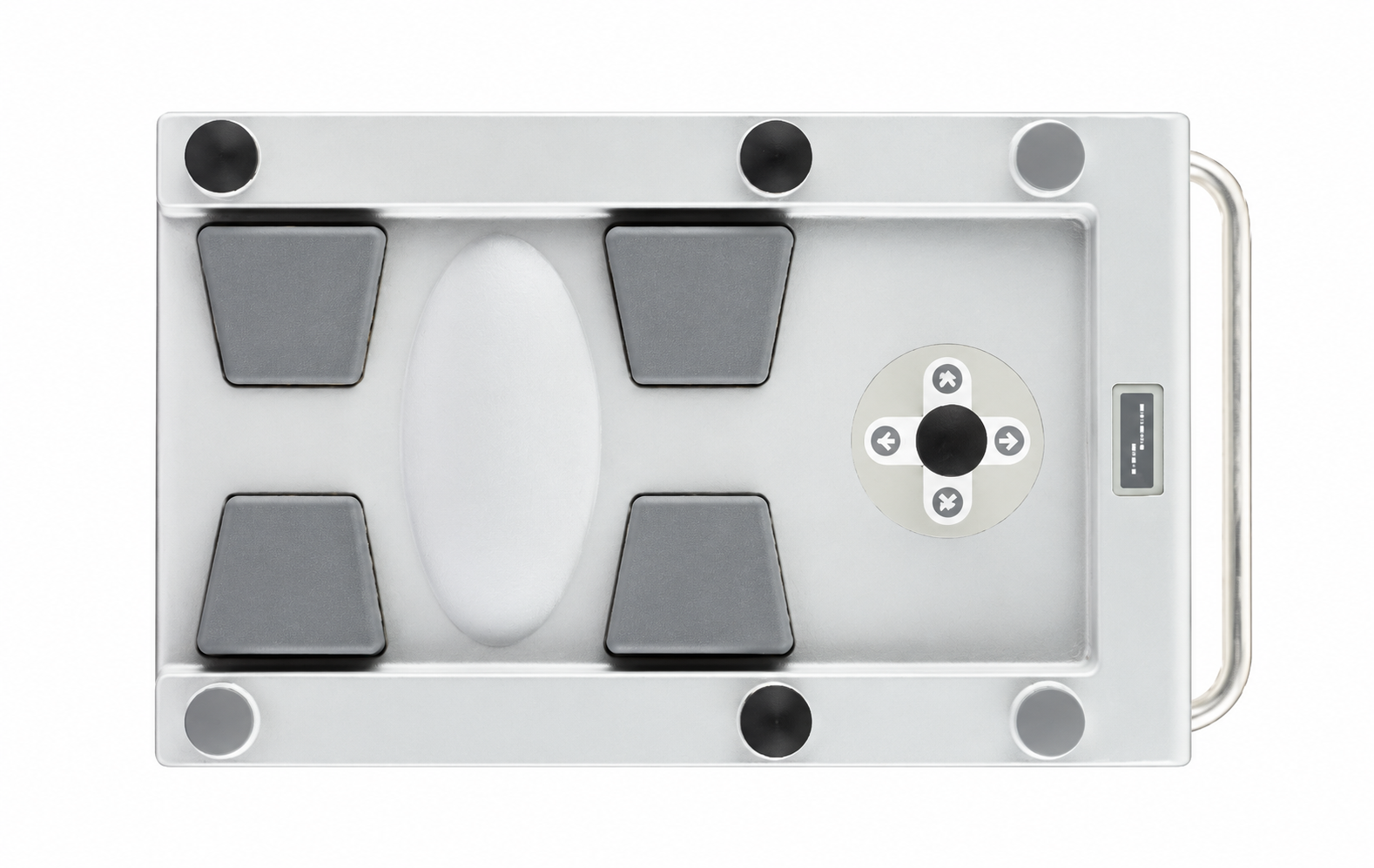
Foot control
Position, focus and illuminate by foot.
The 12-function foot control manages X-Y fine positioning, motorized focus and zoom, brightness and light switching. X-Y movement covers at least 50 × 50 mm, with automatic reset and a movement speed of 2.2 mm/s.
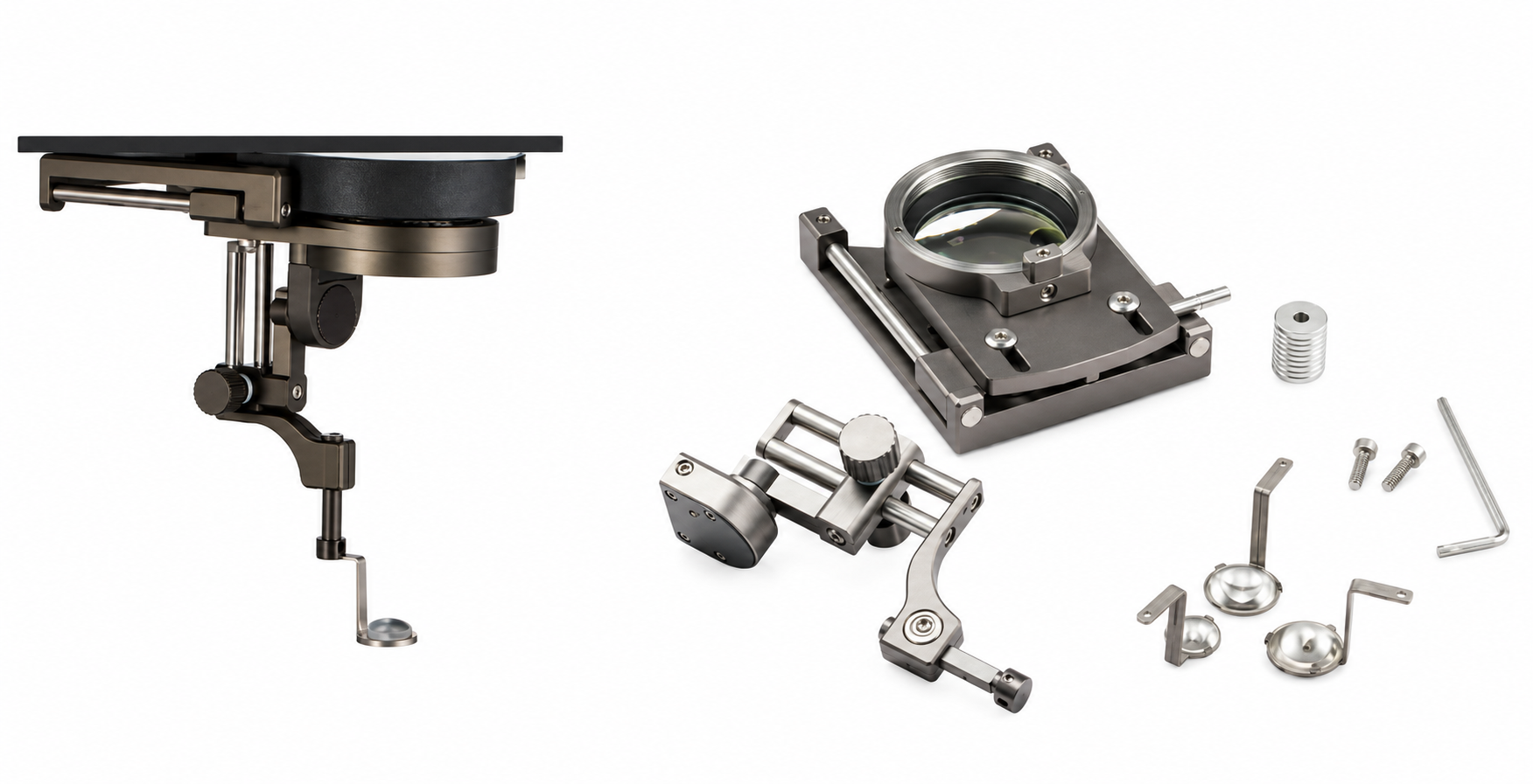
Posterior-segment options
Add posterior-segment viewing.
An optional indirect viewing attachment and optical image inverter extend the microscope configuration for posterior-segment work. The viewing attachment, inverter and mounting components are selected together for the intended setup.
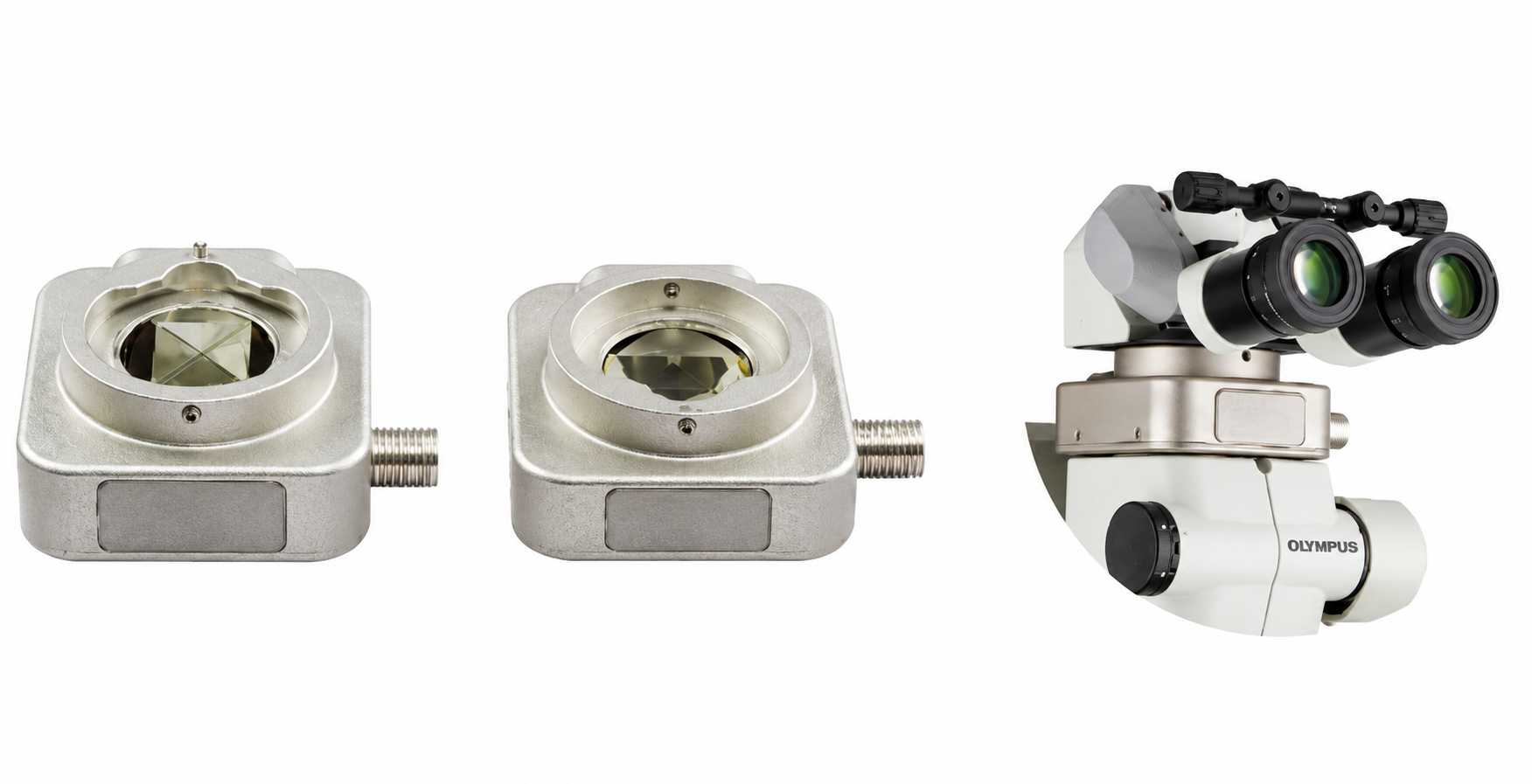
Posterior-segment options
Optical image inversion.
An optional optical image inverter reorients the view produced by the indirect posterior-segment observation system. Specify the viewing attachment, inverter and mounting components together when configuring the microscope.
Product film
See the SM-3000L in motion.
Explore the optics, positioning controls and configuration options in this product demonstration.
Watch the product features, controls and available configurations. Narration and on-screen text are in English.
Configuration questions
Plan the complete setup.
Which posterior-segment components can be specified?
An indirect viewing attachment and an optical image inverter are available as options. Select the viewing attachment, mounting components and inverter together for the intended microscope configuration.
What does the foot control operate?
The 12-function foot control includes X-Y fine positioning, motorized focus, motorized zoom, brightness adjustment and light switching. The control panel also provides eight-level light adjustment.
What recording systems are available?
Optional HD, 4K and 3D stereoscopic imaging systems are available for documentation and teaching. Confirm the camera, adapter, display and recording package with the selected microscope configuration.
Are protective accessories included?
Laser-protective goggles are an optional accessory. The required protection is selected for the actual laser wavelength and application; the final quotation confirms the accessory package.