
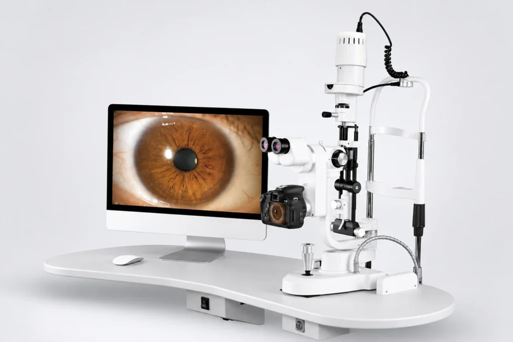
Models: ML-350 / ML-350A
Digital Imaging Configuration Comparison
The ML-350 / ML-350A platform combines slit-lamp observation and LED illumination with a choice of two digital imaging packages. Select the configuration that matches your clinic’s documentation workflow.
| Configuration | ML-350 | ML-350A |
|---|---|---|
| Digital imaging device | Canon EOS 1500D DSLR | Digital CCD module |
| Beam splitter | Integrated beam splitter included | Beam splitter included |
| PC software | Included | Included |
| Computer | Not included | Not included |
| Instrument table | Not included | Not included |
Clear Slit-Lamp Observation
The optical system is designed for clear slit visualization and a wide stereoscopic viewing field. High-point eyepieces support comfortable observation, while sharp slit performance helps the examiner work effectively at lower illumination levels.
What Is a Slit Lamp Imaging System?
A slit lamp, or biomicroscope, combines a stereoscopic microscope with an adjustable slit of light to examine the front of the eye: the eyelids, conjunctiva, cornea, anterior chamber, iris and lens. A slit lamp imaging system adds a beam splitter, a camera and software, so that what the examiner sees can be captured, stored and compared at later visits, shared for referral or shown to the patient. The ML-350 and ML-350A use this set-up, with PC software for image acquisition, processing, patient records, report output and storage.
LED Illumination and Precise Positioning
Warm LED illumination with UV and infrared filtering is designed to reduce heat while supporting long service life. A flexible joystick and quick imaging control help the operator position the instrument and capture findings efficiently.
What Are the Slit Lamp Filters and Settings Used For?
Each filter and beam setting on a slit lamp suits a particular observation. On the ML-350 / ML-350A:
- Cobalt blue filter: used with fluorescein dye to show corneal staining and to assess the tear film and contact-lens fit.
- Red-free filter: makes blood vessels and hemorrhages stand out with higher contrast.
- Neutral-density filter: reduces brightness for patient comfort.
- Heat-absorbing filter: reduces heat from the light path.
- Slit width and length: both are continuously adjustable, width 0-14 mm (a full circle at 14 mm) and length 1-14 mm, from a thin optical section of the cornea to broad diffuse illumination.
- Apertures: from Ø14 mm down to Ø0.2 mm; very small, bright beams are typically used to look for cells and flare in the anterior chamber.
- Magnification: five steps from 6.3X to 40X, low for an overview and high for detail.
Digital Imaging and Documentation Workflow
Both configurations include PC software for image acquisition, processing, patient-record organization, report output and image storage. The ML-350 uses the Canon EOS 1500D DSLR package, while the ML-350A uses a compact digital CCD module.
DSLR or CCD: Which Imaging Configuration?
The two configurations share the same slit-lamp optics, illumination and software; they differ in the camera. The ML-350 uses a Canon EOS 1500D DSLR camera body with an integrated beam splitter, while the ML-350A uses a compact digital CCD module with a beam splitter. Clinics that already work with Canon DSLR equipment may prefer the ML-350, while clinics that want a compact, integrated camera module may choose the ML-350A. In both cases the computer and instrument table are ordered separately.
Ordering Notes
When requesting a quotation, specify either ML-350 with Canon EOS 1500D DSLR or ML-350A with digital CCD module. The corresponding beam splitter and PC software are included; the computer and instrument table are not included. Confirm the final package list, computer compatibility, destination-market power cable, installation, training and service scope in the approved quotation.
What Affects the Price of a Slit Lamp Imaging System?
The price of a slit lamp imaging system depends on the complete working setup rather than the microscope alone. For the ML-350 / ML-350A, the main factors are the imaging configuration (DSLR or CCD), whether a compatible computer and an instrument table are sourced with the order (neither is included), the power cable for your market (the rated supply is 110-220 V, 50/60 Hz), the documentation required, installation and training scope, the order quantity and the shipping terms. Share these details and ENOCHTEC will confirm the configuration and price in a written quotation.
Frequently Asked Questions
What is the difference between ML-350 and ML-350A?
The digital imaging device is the main package difference: ML-350 uses a Canon EOS 1500D DSLR with an integrated beam splitter, while ML-350A uses a digital CCD module with a beam splitter.
Is PC software included?
Yes. PC software is included with both listed digital imaging configurations.
Are the computer and instrument table included?
No. The computer and instrument table are excluded from both configurations.
What magnification does the ML-350 offer?
A five-step revolving drum gives total magnifications of 6.3X, 10X, 16X, 25X and 40X with 12.5X eyepieces.
Does the slit lamp include a cobalt blue filter?
Yes. The ML-350 / ML-350A includes heat-absorbing, neutral-density, red-free and cobalt blue filters.
What light source does the ML-350 use?
A 3 V LED module with luminance of at least 140 klx. The warm LED illumination includes UV and infrared filtering and is designed to reduce heat and support long service life.
View the ophthalmic equipment range, or email ENOCHTEC sales with your market, quantity and preferred imaging configuration.


