
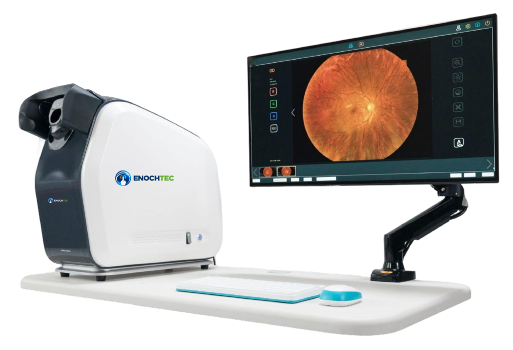
176° True-Color Ultra-Wide-Field Fundus Imaging
The RetiCam 3100 Plus ultra-wide-field fundus camera captures a true-color fundus image of up to 176° in a single exposure. Continuous-spectrum LED illumination renders the retina in natural color, and one-key automatic operation captures both eyes without dilation in routine use, so a wide view of the central and peripheral retina can be documented in one step.
One-Key, Non-Mydriatic Capture of Both Eyes
After patient information is entered, one key starts the examination. The camera searches for the pupil, aligns, adjusts the working distance and focuses automatically, then captures each eye in turn. Gain can be set automatically or manually. The system records at 16 frames per second with an image capture time of 60 ms.
- Automatic pupil search, alignment, working-distance adjustment, focus and capture.
- Automatic or manual gain.
- Working distance: 10 mm ± 2 mm.
Single Image, Two Images and Mosaic
The field of view is quoted from the center of the eyeball:
- Wide field: 176° (tolerance ±5%) in a single image.
- Ultra-wide field: 220° from two images.
- Multi-image mosaic: more than 260°, assembled automatically into one whole-fundus image.
Because ultra-wide-field angles are measured from the eyeball center, they are not directly comparable with the field angles quoted for conventional fundus cameras. Request sample images if you need to compare coverage.
LED True Color and Soft Exposure
Continuous-spectrum LED illumination presents fundus colors close to those seen clinically, and the red, green and blue channels can be viewed separately for contrast review. Optical resolution is 8 μm (tolerance ±7%). Soft exposure technology is designed to reduce pupil constriction after the flash, making consecutive images easier and helping children and patients who are sensitive to light. Maximum flash illumination is below 130,000 lx.
Image Review and Analysis Software
Images are reviewed on a 27-inch color monitor with 2560 × 1440 resolution. The software adjusts brightness, gamma, contrast and RGB gain, and provides tools to measure distances and lesion area and to estimate the cup-to-disc ratio. Measurement methods, validation and software version should be confirmed for the order.
RetiCam 3100 Plus or RetiCam 3100?
Both are automatic non-mydriatic fundus cameras from ENOCHTEC. They differ in field of view and illumination.
| Item | RetiCam 3100 Plus | RetiCam 3100 |
|---|---|---|
| Single-image field | 176° ultra-wide field (from eyeball center) | 50° |
| Wider coverage | 220° from two images; over 260° by mosaic | Up to 135° from a nine-image mosaic |
| Illumination / camera | Continuous-spectrum LED, true color | SLR camera photography |
| Working distance | 10 mm ± 2 mm | 13 mm |
- Choose the RetiCam 3100 Plus when a wide single-image view of the peripheral retina is useful, for example in retina clinics and hospital departments that review peripheral findings.
- Choose the RetiCam 3100 for standard 50° posterior-pole documentation with mosaic, DICOM and a customizable AI interface.
RetiCam 3100 Plus Questions Before Ordering
Is 176° comparable with the field angle of a conventional fundus camera?
Not directly. The 176° figure is measured from the center of the eyeball, while conventional fundus cameras usually quote the field differently. Compare sample images rather than angles alone.
Does every examination avoid dilation?
The camera is designed for non-mydriatic capture with automatic pupil search. The clinician decides the appropriate preparation and whether an image is adequate for the intended examination.
What power supply does it use?
The listed supply is 220 V with 200 W power. Confirm the destination-market voltage, plug and documentation in the quotation.
What is included in the package?
Confirm the imaging unit, monitor and arm, computer, table, software, accessories, training and service scope in the quotation.
Request a RetiCam 3100 Plus Configuration
Contact ENOCHTEC sales with your country, quantity, intended examination setting and image-management requirements. Confirm the current model-specific specification, workstation, accessories, software, training, service and destination-market documentation in the quotation. Specifications and design are subject to change.