
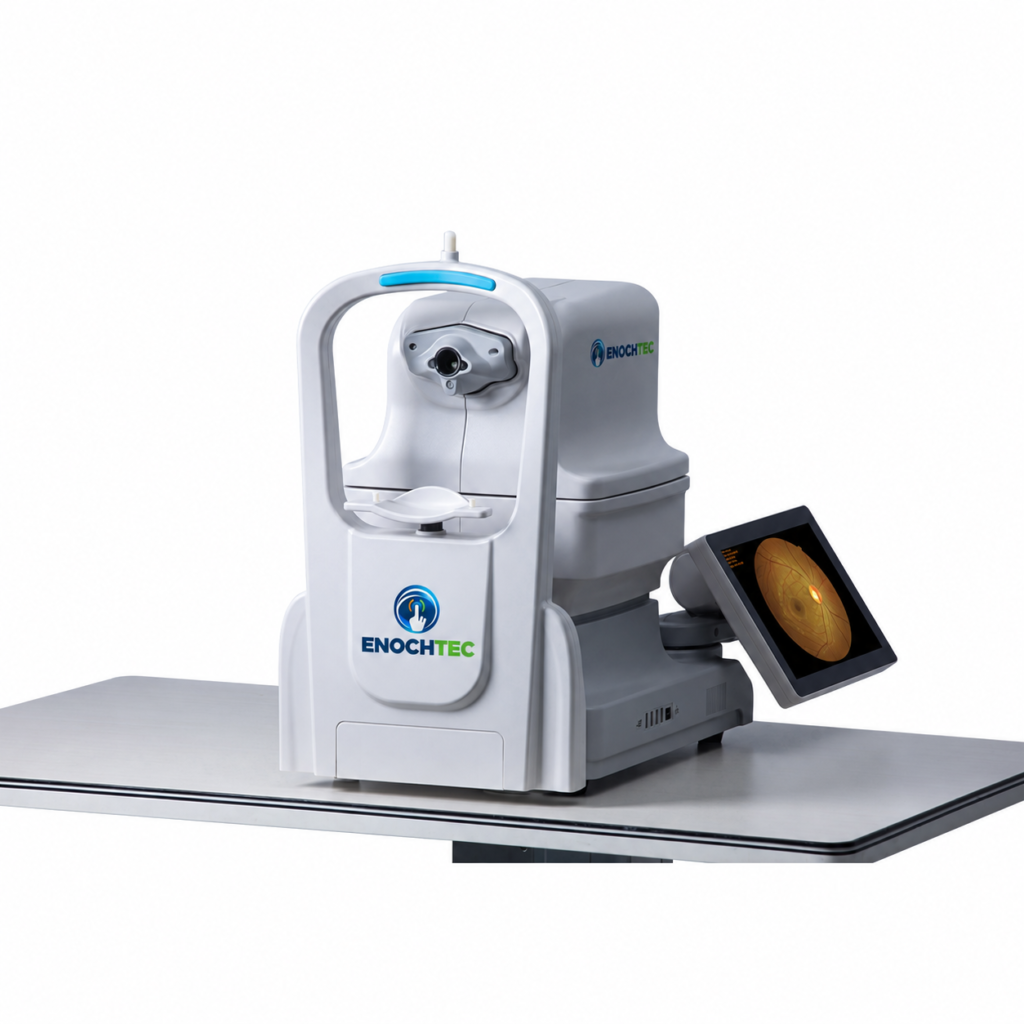
One-Key Automatic Fundus Imaging for Small Pupils
The Kestrel 3100m non-mydriatic automatic fundus camera captures 50° color fundus images of both eyes with one key. The camera adjusts the chinrest, searches for the pupil, aligns, focuses and fires automatically, and a small-pupil mode supports imaging through pupils as small as 2.8 mm.
Why Choose the Kestrel 3100m
- Fully automatic one-key capture: the chinrest moves up and down automatically, and the camera searches, aligns, focuses and shoots by itself.
- Fast examinations: both eyes in about 15–20 seconds; SYSEYE describes it as the world’s fastest automatic fundus camera.
- Built-in computer: an all-in-one design that is easy to install and takes up little space.
- Small pupils: fundus images through pupils as small as 2.8 mm, where many automatic fundus cameras need 3.3–3.8 mm.
- Cost-effective: automatic small-pupil fundus imaging at a competitive price for clinics and screening programs.
Fully Automatic Operation with a Dual-Camera System
After patient information is entered, one click starts the sequence: automatic chinrest adjustment, pupil searching, alignment, focusing and capture, followed by the second eye. A dual-camera system locates the pupil quickly for fast and accurate autofocus. Manual mode is available when automatic capture is not suitable or a particular area needs to be photographed, with alignment, focus, flash and diopter compensation controlled on the touchscreen.
- Automatic pupil searching, chinrest adjustment, focusing and shooting.
- Automatic and manual focus, exposure and operation.
- Soft lighting reduces the pupil constriction caused by illumination, so satisfactory images can be obtained even in two consecutive exposures.
Variable Fixation and 135° Mosaic
Besides the nine preset internal fixation positions, the operator can set any point on the touchscreen as the fixation point to photograph a specific retinal area. The mosaic function combines nine 50° images into a composite fundus image of up to 135°; this is a multi-image composite, not a single exposure.
All-in-One, Space-Saving Design
The camera, computer and rotatable touchscreen monitor are integrated in one device, which keeps the examination area compact. An additional external monitor can be connected for teaching or for the doctor’s review.
Image Software, Storage and Transmission
The built-in software adjusts brightness, gamma, contrast and RGB, compares images side by side and measures lesion area and cup-to-disc ratio. USB and LAN interfaces allow flexible storage and sharing: DICOM 3.0 for hospital PACS connection, LAN sharing for real-time viewing on other computers, USB storage expansion and remote transmission by FTP. A customizable AI port is provided; AI reading software and its regulatory status are not assumed included.
Upgraded Imaging Configuration
The manufacturer’s 2026 brochure presents red-free, fluorescein angiography (FFA) and fundus autofluorescence (FAF) imaging as an upgraded Kestrel 3100m configuration. Confirm the configuration supplied, and its specifications, in the quotation.
Kestrel 3100m or RetiCam 3100?
Both are SYSEYE automatic non-mydriatic fundus cameras with a 50° field and a 135° nine-position mosaic.
| Item | Kestrel 3100m | RetiCam 3100 |
|---|---|---|
| Image resolution | 5 MP (SLR camera) | 24 MP (SLR camera) |
| Minimum pupil | ≥2.8 mm | ≥3.3 mm |
| Working distance | 13 mm | 35 mm |
| Fixation | Internal, with operator-defined points | External / internal |
| Versions | One model | Standard, FFA and FAF versions |
- Choose the Kestrel 3100m for compact clinics and screening sites that see many patients with small pupils.
- Choose the RetiCam 3100 when higher image resolution or a dedicated FFA or FAF version is needed.
Kestrel 3100m Questions Before Ordering
Can patients with small pupils be imaged without dilation?
The small-pupil mode supports pupils as small as 2.8 mm, and a mydriatic mode is also listed. The clinician decides on patient preparation and whether an image is adequate for the intended examination.
Is angiography included?
Red-free, FFA and FAF imaging are presented by the manufacturer as an upgraded configuration. Confirm the configuration and its specifications in the quotation.
Can it connect to our PACS?
DICOM 3.0 is supported for PACS connection, together with LAN sharing, USB storage and FTP transfer. Confirm compatibility with the receiving system before installation.
Request a Kestrel 3100m Configuration
Contact the ENOCHTEC sales team with your country, quantity, clinical setting and image-management requirements. Confirm the current model specification, configuration, software, accessories, training, service and destination-market documentation in the quotation. Specifications and design are subject to change.