
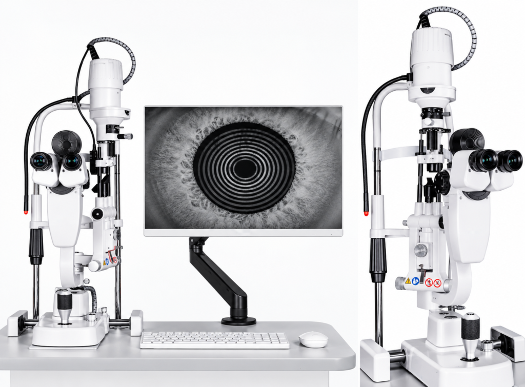
Dry Eye Analyzer Built on a Slit Lamp Platform
The SLR-3000 dry eye analyzer brings non-invasive tear film, lipid layer and meibomian gland assessment to a slit lamp platform. A rotating 25-ring Placido module sits in front of the microscope, so the operator can move between dry eye examination and anterior segment imaging without inserting or removing an accessory. The system records video of tear film stability, incomplete blinking and lipid layer behavior, and captures images of the tear meniscus, lipid layer and meibomian gland openings.
Non-Invasive Tear Film Assessment
Tear stability evaluation uses the projected Placido rings to recognize tear film break-up automatically and without touching the eye, showing both the stability and the regularity of the tear film. Non-invasive break-up time can be analyzed automatically or reviewed manually. Tear meniscus height measurement lets the examiner assess the height, regularity and shape of the tear meniscus, also with automatic or manual analysis.
Lipid Layer, Meibography and Gland Openings
Static and dynamic lipid layer images are shown on the same screen, so the examiner can follow how the lipid layer changes instead of comparing separate captures. Meibography provides an enhanced, low-distortion view of the meibomian glands, making gland dropout easier to recognize. Gland opening evaluation captures the overall shape of the openings along the lid margin to help assess blockage.
Blink, Redness and Corneal Staining
The SLR-3000 analyzes the incomplete blink rate automatically. Ocular redness classification displays the hierarchy of ocular blood vessels, the overall degree of congestion and the position of dilated vessels. For corneal staining, the built-in yellow filter is used with cobalt blue illumination to make corneal defects visible.
Optional Mite Detection
Mite detection is an optional mode. It uses high-magnification imaging to examine eyelash follicles for Demodex folliculorum, supporting the assessment of lipid-related dry eye, and is presented in the source brochure as an imaging alternative to conventional biomicroscopy that can shorten patient waiting time. Specify this option in your inquiry if it is required.
Dry Eye Progress Trends and Reports
Results from each visit can be recorded to build a dry eye progress trend chart. Following several indicators over time helps clinicians evaluate severity and progression during treatment and plan individualized care. One report can combine multiple examination results, and reports can be printed as a comprehensive summary or item by item, using single or multiple customized layouts.
Anterior Segment Imaging
After dry eye testing, the same instrument is used for anterior segment examination and imaging. The source brochure describes high-definition observation with realistic color reproduction, so captured images reflect the actual appearance of the eye. Optical, slit, filter and image-sensor data are listed in the specification table further down this page.
Choosing the Right ENOCHTEC Slit Lamp
Choose the SLR-3000 when the main requirement is dry eye and ocular surface analysis. For general slit lamp documentation with a camera package, compare the ML-350 / ML-350A digital slit lamp microscope; for bedside or mobile examinations, see the PSL-4961 handheld slit lamp. All related models are grouped under slit lamp examination.
SLR-3000 Frequently Asked Questions
What does the SLR-3000 dry eye analyzer evaluate?
It supports non-invasive tear film break-up time, tear meniscus height, static and dynamic lipid layer evaluation, meibography, gland opening evaluation, incomplete blink rate, ocular redness classification, corneal staining and dry eye progress trends, in addition to anterior segment imaging.
Does the Placido module have to be removed for routine slit lamp work?
No. The 25-ring Placido module rotates into position, so it does not need to be inserted or removed when switching between dry eye and anterior segment examination.
Is mite detection included as standard?
No. Mite detection is an optional configuration and should be specified when you request a quotation.
Are a computer, monitor, printer, instrument table and software license included?
The source material does not state this. Whether each of these items is supplied is confirmed in the quotation. Items shown in the product images are for illustration only.
Review more ophthalmic diagnostic and imaging equipment, or contact ENOCHTEC to confirm the SLR-3000 configuration and supply scope.

