IVS-201A / IVS-201B Hidden LED Array Visual Field Analyzer
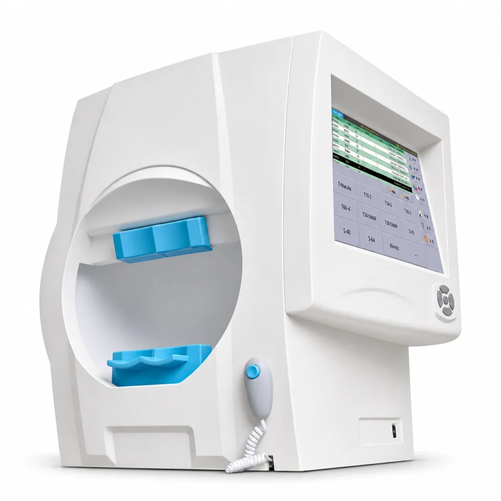
The ENOCHTEC IVS series is an automated visual field analyzer with a hidden LED array for standard white-on-white threshold and screening perimetry. The IVS-201A adds blue-yellow perimetry (SWAP), custom test programs, glaucoma progression analysis and DICOM networking; the IVS-201B covers the core glaucoma, macular and neurological programs at a lower configuration. Both run on a dedicated embedded system with a 15-inch touchscreen.
IVS-201A or IVS-201B?
Both models share the same bowl, stimulus system, threshold strategies, fixation control and basic analysis. The differences are:
| Function | IVS-201A | IVS-201B |
|---|---|---|
| White-on-white threshold and screening programs | ✓ | ✓ |
| HISA, Full Threshold, Fast Ladder and Standard Threshold strategies | ✓ | ✓ |
| Gaze tracking, blink control, pupil measurement | ✓ | ✓ |
| VFI, GHT, single field analysis, serial field overview | ✓ | ✓ |
| Blue-yellow perimetry (T24-SWAP, T30-SWAP) and SWAP analysis | ✓ | — |
| Custom test programs | ✓ | — |
| Blue-stimulus colored perimetry and specialty patterns | ✓ | — |
| Glaucoma progression analysis | ✓ | — |
| Networking and DICOM support | ✓ | — |
Standard White-on-White Perimetry
The IVS series uses a white stimulus on a 31.5 asb (10 cd/m²) background, the illumination standard used by mainstream projection perimeters, with Goldmann III stimuli from 0.08 to 10,000 asb (0–50 dB) at a 30 cm test distance. Threshold programs include T30-2, T24-2 and T10-2 for glaucoma, T-Macula for macular function, T60-4 for the peripheral field and T-NS for the nasal step. Screening programs cover the central and full field, including S-Armaly, with two-zone, three-zone and quantified-defect strategies. The maximum temporal range is 90°.
HISA Threshold Strategy
The HISA strategy estimates the starting threshold of each point from neighbouring results and age-matched normal values, skips unnecessary presentations, adapts the stimulus interval to the patient’s response speed and retests points whose results look unreliable. This shortens examination time for both fast and slow responders without changing the test pattern.
Blue-Yellow Perimetry (SWAP) on the IVS-201A
Short-wavelength automated perimetry tests the blue-cone pathway with a 440 nm blue stimulus on a bright 100 cd/m² yellow background. Studies suggest that SWAP can reveal glaucomatous field loss in some patients earlier than standard white-on-white perimetry. The IVS-201A runs T24-SWAP and T30-SWAP with a Goldmann III stimulus and the HISA-SWAP algorithm, so a blue-yellow test takes about as long as a standard test.
SWAP results show more variability than standard perimetry and are affected by yellowing of the crystalline lens, so SWAP is used alongside standard perimetry and structural imaging such as the OCT-500 spectral-domain OCT, not as a replacement. Interpretation remains the responsibility of the examining clinician.
Fixation Control and Reliability
Gaze tracking monitors pupil position throughout the test: the system alerts the patient when fixation drifts, ignores stimuli presented during an eye movement, pauses the test if the shift persists and can move the chin rest to re-centre the pupil. Stimuli missed during a blink are repeated automatically. Heijl-Krakau blind-spot monitoring, infrared video, false-positive and false-negative catch trials and a gaze-tracking curve help the clinician judge test reliability, and the pupil diameter is measured and printed on the report.
Analysis and HFA-Style Reports
Reports show numeric values, grayscale, total and pattern deviation with probability plots, the Visual Field Index (VFI), pattern standard deviation (PSD) and the Glaucoma Hemifield Test (GHT). The familiar HFA-style layout makes results easy to compare with other mainstream perimeters. On the IVS-201A, glaucoma progression analysis separates significant change from test-to-test variability across a series of follow-up fields.
Dedicated System, Storage and Networking
Instead of a general-purpose PC, the IVS series uses an embedded ARM computer with a dedicated operating system, which avoids common PC viruses and moving parts. More than one million test results are stored on at least 32 GB of solid-state memory, with backup to a flash disk, portable hard disk or network. The IVS-201A supports Ethernet networking and DICOM for connection to compatible EMR and image-management systems. The 15-inch touchscreen is angled toward the operator and shielded from the patient.
IVS Series vs BIO-1000 Plus
| IVS-201A | IVS-201B | BIO-1000 Plus | |
|---|---|---|---|
| Background / test distance | 10 cd/m², 30 cm | 10 cd/m², 30 cm | 10 cd/m², 300 mm |
| Central threshold programs | 30-2, 24-2, 10-2, macula | 30-2, 24-2, 10-2, macula | 30-2, 24-2, 10-2, macular, nasal ladder |
| Blue-yellow perimetry (SWAP) | Yes | No | Not listed |
| Operator interface | Built-in 15-inch touchscreen | Built-in 15-inch touchscreen | Computer-based operation |
| DICOM networking | Yes | No | Confirm with quotation |
| Power supply | 100–240 V, 50–60 Hz | 100–240 V, 50–60 Hz | 220 V, 50 Hz |
Choose the BIO-1000 Plus for standard static threshold and screening perimetry; the IVS-201A when blue-yellow perimetry, progression analysis or DICOM integration is required; and the IVS-201B for core programs on a self-contained touchscreen system that works on 110 V or 220 V mains.
Where the IVS Fits in a Glaucoma Workup
- Intraocular pressure — with the ST-1000P non-contact tonometer or the RT-500 rebound tonometer.
- Corneal thickness — with the MD-1000P ultrasonic pachymeter, as a reference for IOP interpretation.
- Structure — optic nerve and RNFL imaging with the OCT-500.
- Function — visual field testing and follow-up with the IVS series.
Request a glaucoma equipment package quote.
What Should Buyers Confirm Before Ordering?
Confirm the model (IVS-201A or IVS-201B), whether SWAP, progression analysis or DICOM is needed, the optional foot-pedal responder, keyboard and mouse, the interface and report language, installation space (56 × 49 × 60 cm, 30 kg) and the documentation required in the destination market. See the Visual Field Analysis & Perimetry range for related equipment. The final supply configuration is confirmed in the written quotation.
What Affects the Price of a Visual Field Analyzer?
The price depends mainly on the model — the IVS-201A with SWAP, progression analysis and DICOM, or the IVS-201B — plus optional accessories, networking requirements, language and documentation for the destination market, and shipping terms. Including these points in your enquiry allows an accurate, comparable quotation.
Frequently Asked Questions
What is the difference between the IVS-201A and IVS-201B?
Both share the same hardware and core programs. The IVS-201A adds blue-yellow perimetry (T24-SWAP and T30-SWAP), custom programs, blue-stimulus and specialty tests, glaucoma progression analysis, networking and DICOM support.
What is SWAP perimetry?
Short-wavelength automated perimetry presents blue stimuli on a yellow background to test the blue-cone pathway. It is used alongside standard white-on-white perimetry in glaucoma assessment.
Which visual field programs are included?
Threshold programs T30-2, T24-2, T10-2, T-Macula, T60-4 and T-NS, plus central and full-field screening programs such as S-Armaly. The IVS-201A adds T24-SWAP, T30-SWAP and custom programs.
How does the IVS control fixation?
Gaze tracking, infrared video, Heijl-Krakau blind-spot monitoring and blink control; stimuli presented during eye movements or blinks are discarded or repeated.
Does the IVS need a separate computer?
No. It runs on a built-in embedded system with a 15-inch touchscreen; a keyboard and mouse are optional.
What power supply does it need?
100–240 V, 50–60 Hz, about 150 W, so the same unit works on 110 V and 220 V mains.