
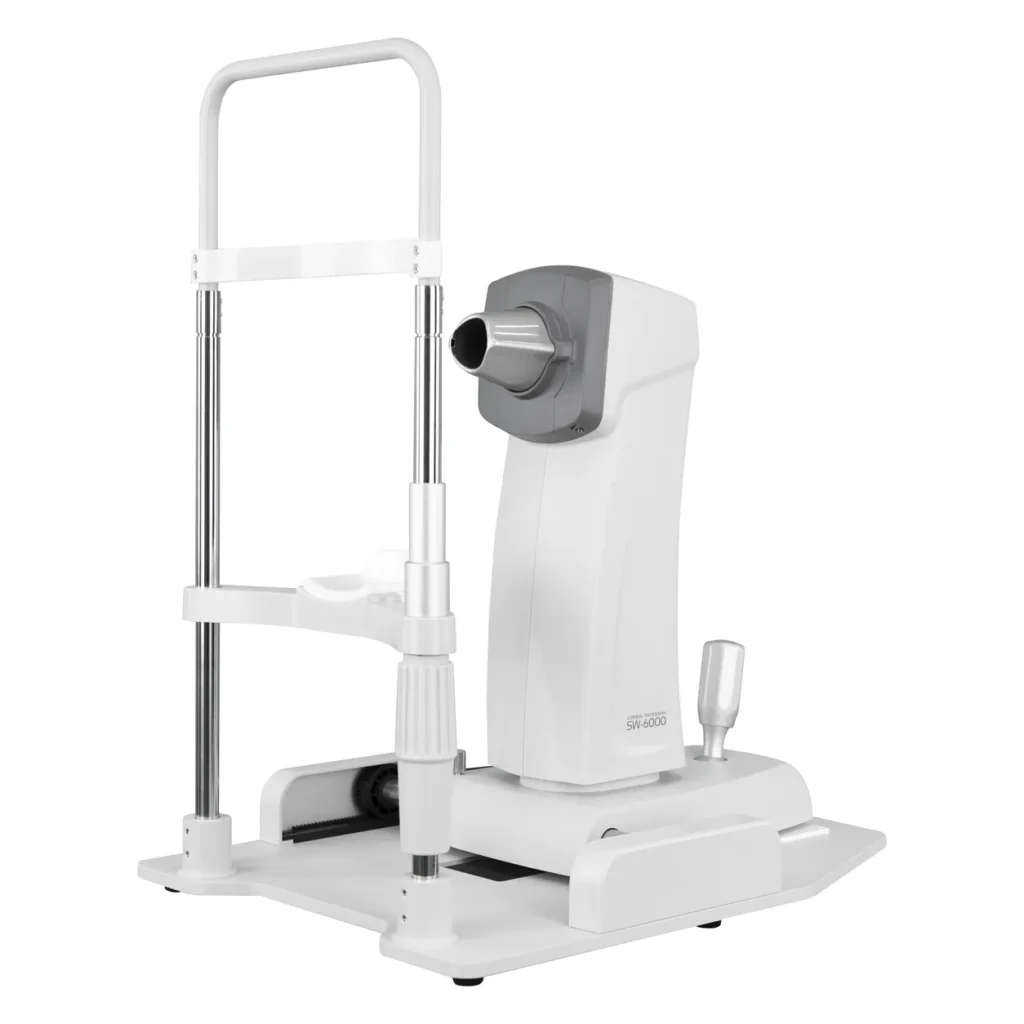
SW-6000 Placido Corneal Topographer
The SW-6000 corneal topographer maps the front surface of the cornea from a single Placido-ring capture. Its 31-ring disc gives 7,936 measured points across a 10.91 mm zone, and the software turns them into axial, tangential and elevation maps, simulated keratometry and keratoconus screening indices. Clinics use it for refractive surgery screening, keratoconus follow-up, orthokeratology and rigid contact lens fitting, and general assessment of the corneal surface.
Corneal Maps and Indices
Each examination can be shown as single maps or in combined layouts. Results support the clinician’s assessment; the maps and indices do not make a diagnosis on their own.
- Axial and tangential curvature maps in dioptres or millimetres.
- Elevation map against a best-fit sphere, with the fit radius shown.
- Keratoscope image of the reflected Placido rings, useful for checking tear film and capture quality.
- Simulated K in 3, 5 and 7 mm zones: steep and flat meridians, axes and corneal astigmatism.
- Shape factor, eccentricity and a meridian profile graph for each map.
- Keratoconus view with screening indices and a 3D surface view.
How Placido Topography Works
The SW-6000 projects 31 concentric rings onto the tear film and records their reflection with a camera. The spacing and shape of the reflected rings change with corneal curvature: steeper areas compress the rings and flatter areas spread them. The software measures 7,936 points along the rings and reconstructs the anterior corneal surface over 10.91 mm, with a radius range of 5.5–10.0 mm and an accuracy of ±0.02 mm. Placido topography measures the front surface only; posterior surface and thickness maps require a Scheimpflug or OCT-based tomographer.
Capture and Patient Positioning
The patient rests on the chinrest and forehead rest and fixates on the target inside the cone. The operator centres and focuses the image with the joystick, guided by on-screen distance prompts such as “Too Far”, and the joystick moves the measuring head 86 mm laterally, 40 mm fore–aft and 30 mm vertically; the chinrest adjusts by 50 mm. A stable tear film and good lid opening give the most complete ring image, so contact lens wearers should follow the clinic’s usual lens-removal interval before topography.
Software Views and Patient Records
Examinations are stored by patient, eye and date. The software offers Single, Compare, Combine, 3-map, 4-map, Numerical, Keratoconus and Double-eyes layouts, so two examinations or both eyes can be reviewed side by side. Map options such as colour map, rings, grid, polar overlay, section and numeric readout can be switched on or off, and the keratometric index can be set (for example 1.3375).
Contact Lens Fitting
The contact lens module suggests starting parameters for rigid gas-permeable and orthokeratology lenses from the measured topography. Final lens selection remains with the fitter, based on trial lenses and follow-up examinations.
Workstation Setup
The SW-6000 measuring unit connects to a computer running the analysis software. A computer, monitor, keyboard, colour printer, printer shelf and height-adjustable instrument table can be supplied as a complete workstation; these items are quoted separately and confirmed for each project.
SW-6000 Overview Video
SW-6000 or an Auto Refractor Keratometer?
An auto refractor keratometer such as the ARK-8900 measures refraction and central corneal curvature at a few points about 3 mm apart. The SW-6000 maps 7,936 points across 10.91 mm, so it shows irregular, peripheral and post-surgical corneal shape that K readings cannot. Many clinics use both: the ARK for every patient and topography for screening and lens fitting. For a detailed comparison, see Keratometer vs Corneal Topographer.
Applications
The SW-6000 supports refractive surgery screening, keratoconus follow-up, orthokeratology and rigid lens fitting, documentation after refractive or corneal surgery, and anterior corneal assessment as supporting information before cataract surgery. Typical users include ophthalmology clinics, refractive surgery centres, optometry and contact lens practices, and distributors of ophthalmic equipment.
Frequently Asked Questions
How is the SW-6000 different from a keratometer?
A keratometer reads curvature at a few points in the central cornea. The SW-6000 measures 7,936 points across 10.91 mm and shows the whole anterior surface as a colour map, including irregular and peripheral shape.
Does the SW-6000 measure the back of the cornea or corneal thickness?
No. As a Placido topographer it maps the front corneal surface only. Posterior elevation and pachymetry maps need a Scheimpflug or OCT-based tomographer.
Can the SW-6000 diagnose keratoconus?
It provides screening indices and colour-coded maps that flag suspicious patterns. The diagnosis remains the clinician’s, based on the maps, the indices and the full examination.
Is it suitable for orthokeratology fitting?
Yes. The contact lens module suggests initial lens parameters from the measured topography, and follow-up maps show how the cornea has responded.
Is the computer included?
The computer, monitor, keyboard, printer and instrument table are quoted separately, so the workstation can match local requirements or equipment the clinic already has.