
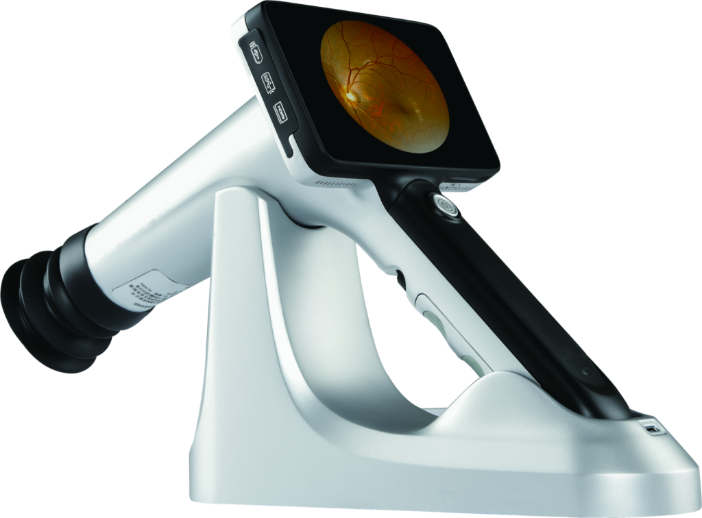
The CFC-X portable fundus camera is a handheld, non-mydriatic retinal imaging device designed for screening and documentation workflows in clinics, optical practices, community programs and mobile eye-care settings. Its compact form combines a 50° field of view, 18 MP sensor resolution, automatic and manual focus, and an integrated 4.7-inch anti-glare touchscreen.
CFC-X Imaging Workflow Video
Portable Fundus Camera for Flexible Workflows
Traditional tabletop fundus cameras can be difficult to move between examination rooms or take into outreach environments. The CFC-X is designed to support retinal photography where mobility matters. At less than 600 g, the camera can be used as a handheld unit and returned to its charging cradle between examinations. An optional portable stabilization support is also available, helping operators maintain a consistent working position during image capture.
50° Non-Mydriatic Fundus Imaging
The camera uses infrared and white LED illumination for non-mydriatic imaging. The minimum pupil diameter is 2.5 mm and the working distance is 24 mm. The single-image field of view is 50°. Multi-point fixation can guide additional captures for broader retinal documentation; coverage reaches up to 90° when multiple fixation positions are used. This wider coverage figure should not be interpreted as the field of view of one exposure.
Automatic and Manual Focus
Both automatic and manual focus modes are available, allowing the operator to choose a faster screening workflow or make fine adjustments when required. Artificial fixation points help guide gaze direction, while the integrated display allows images and patient records to be reviewed directly on the device. The four-core processor and GPU-based hardware are intended to support image processing and integration with compatible third-party AI software.
Connectivity and Data Handling
The CFC-X supports USB 3.0, Wi-Fi, Bluetooth, SD card and Type-C connections, together with 4G and NFC functions. These interfaces provide options for transferring retinal photographs and connecting the camera to an established documentation workflow. Exact software functions, file formats, DICOM availability, AI integration and regional configurations should be confirmed before ordering.
Sample Fundus Images and AI-Assisted Report
The images and report below are samples from the CFC-X, shown with an example report produced by compatible third-party AI software. They are provided for illustration only. Availability of AI analysis depends on the configuration and local regulatory requirements and should be confirmed before ordering. AI output supports, but does not replace, clinical interpretation.
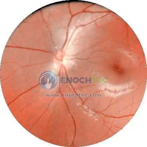
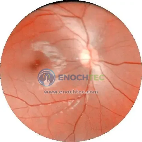
- Name
- Sample patient
- Gender / Age
- Male / 66
- Hypertension
- No
- Diabetes
- No
- Recent vision (left / right)
- Denial of decreased vision
- History of eye disease
- Deny
- (OS) Left eye
- Good image quality (high image readability)
- (OD) Right eye
- Good image quality (high image readability)
- (OS) Left eye
- No diabetic retinopathy
- (OD) Right eye
- No diabetic retinopathy
Video: How the AI-Assisted Report Is Generated
Designed for Clinic, Community and Mobile Screening
Potential workflows include ophthalmology and optometry examinations, diabetic retinopathy screening programs, community eye-health projects, bedside retinal documentation and mobile screening. The device is intended to support image acquisition and review by trained professionals; it does not replace clinical interpretation or diagnosis.
Quick Imaging Workflow
A typical CFC-X capture sequence follows six steps on the touchscreen:
- Power on and rough focus. Press the power button to open the live preview. Holding the handle with one hand and supporting the lens barrel with the other, focus on a scene about 3 m away so the preview is sharp before approaching the patient.
- Choose the imaging mode. Select color or red-free imaging, then automatic (AF) or manual (MF) shooting.
- Create a case. Tap the new-case icon, enter the name, case ID and date of birth, and save the record.
- Select the eye. Tap the left-eye or right-eye icon so images are filed to the correct side.
- Switch on the fixation light. Use automatic or manual fixation as required; in manual mode, tap a fixation point to light it (green means active). The right fixation light is normally used for the left eye, and the left fixation light for the right eye.
- Align and capture. Bring the camera to the working distance until the alignment indicator is clear, center the optic disc or macula in the live view and capture.
Image Quality Tips for Handheld Fundus Photography
Consistent handheld images depend as much on preparation and technique as on the camera. Seat the patient comfortably facing forward with the head stable, and ask them to keep the eye wide open and avoid blinking during capture. Dimming the room light helps the pupil stay larger for non-mydriatic imaging; a few blinks beforehand can reduce tear-film reflections.
Hold the camera with both hands, keep it level with the patient’s eye and rest your elbows lightly on a table or against your body. Slow, controlled adjustments work better than sudden movements. For general screening, macula-centered images are usually preferred; optic disc–centered images are needed for glaucoma-focused workflows. Taking two to three images per eye helps ensure at least one high-quality image is available.
- Reflection or glare: re-adjust the angle or reduce ambient light.
- Shadow or vignetting: keep the correct working distance and center the pupil.
- Blurred image: check focus and make sure the patient’s eye is not moving.
- Small-pupil artifacts: dim the room and ask the patient to keep the eye wide open.
Battery and Daily Care
The CFC-X is designed for more than eight hours of operation per charge and recharges in less than two hours. To avoid running the battery flat, charge the camera every 3–5 days, and do not leave a single charging session longer than eight hours. The camera cannot be used while it is charging; unplug the charging cable before starting an examination.
Configuration and Accessories
The CFC-X comprises the handheld camera, charging cradle, portable stabilization support and protective carrying case. Included accessories can vary by quotation and market. ENOCHTEC can help confirm the required configuration, compatible software and documentation for a planned screening or clinic workflow.
Request a CFC-X Configuration
Contact ENOCHTEC for current configuration details, accessory availability, lead time and a quotation for the CFC-X handheld non-mydriatic fundus camera.